

Invasive breast carcinoma - IARC
Invasive breast carcinoma - IARC
Invasive breast carcinoma - IARC

You also want an ePaper? Increase the reach of your titles
YUMPU automatically turns print PDFs into web optimized ePapers that Google loves.
Clinical features<br />
Patients range in age from 15-81 years,<br />
and most present with a unilateral adnexal<br />
mass. Ultrasound studies may show<br />
an ill defined mass {637}.<br />
Macroscopy<br />
These predominantly solid tumours<br />
range from 0.5-18 cm in diameter. The<br />
sectioned surface may contain variably<br />
sized cysts and is yellow-tan to greywhite<br />
{2877}. The tumour is firm to rubbery<br />
and occasionally may have areas of<br />
haemorrhage and necrosis.<br />
Tumour spread and staging<br />
Tumour implants may be present at the<br />
time of diagnosis and indicate an<br />
aggressive tumour {637,2653}.<br />
Histopathology<br />
The tumour shows a variable admixture<br />
of diffuse, solid and sieve-like cystic<br />
areas, with the solid pattern dominating<br />
in the majority of cases. The diffuse, solid<br />
areas show a compact proliferation of<br />
ovoid to spindle-shaped cells reflecting<br />
closed tubules bound by a basement<br />
membrane and separated by variable<br />
amounts of fibrous stroma or none at all.<br />
The round to ovoid nuclei may show<br />
indentations. The hollow tubules have a<br />
retiform or sertoliform appearance. When<br />
the closed tubules dominate, the lesion<br />
resembles a mesenchymal tumour; a<br />
PAS or reticulin stain helps unmask the<br />
tubular pattern. The cells lining the<br />
tubules are cuboidal to low columnar<br />
with a minimal amount of eosinophilic<br />
cytoplasm and round to spindle-shaped,<br />
uniform nuclei. Sieve-like areas display<br />
clusters of variably sized cysts lined by<br />
attenuated cells. Most cases do not show<br />
atypia or mitotic figures.<br />
Immunoprofile<br />
The tumour cells are positive for most<br />
cytokeratins and vimentin and are often<br />
positive for calretinin (91%), inhibin<br />
(68%) and CD10 {2110}. They are usually<br />
negative for epithelial membrane antigen,<br />
estrogen receptor (ER) and progesterone<br />
receptor (PR) and are negative for<br />
cytokeratin 20, 34betaE12 and glutathione<br />
S-transferase {682,2926}.<br />
Cytometry<br />
The ploidy of a metastatic tumour was<br />
assessed and found to be diploid {2653}.<br />
Electron microscopy<br />
At the ultrastructural level, the tubules<br />
are surrounded by basal lamina and<br />
lined by cells with complex interdigitations,<br />
desmosomes and/or tight junctions<br />
and a few microvilli along the luminal border;<br />
no cilia are identifiable {670}. The<br />
cytoplasmic organelles are not distinctive<br />
and include lysosomes, a small<br />
amount of smooth endoplasmic reticulum<br />
and a few lipid droplets.<br />
Differential diagnosis<br />
The main tumours in the differential diagnosis<br />
are Sertoli cell tumour, Sert o l i -<br />
Leydig cell tumour, and well differentiated<br />
endometrioid <strong>carcinoma</strong>. The presence<br />
of a sieve-like pattern and the<br />
absence of Leydig cells help distinguish<br />
wolffian tumours from all these lesions.<br />
The absence of immunoreactivity with<br />
either ER or PR also would distinguish<br />
wolffian tumours from well differentiated<br />
endometrioid <strong>carcinoma</strong>s; the latter are<br />
invariably positive for ER and PR; howeve<br />
r, positive immunostaining does not<br />
exclude the possibility of a wolff i a n<br />
tumour {682}.<br />
Prognosis and predictive factors<br />
The tumour stage as well as cytological<br />
atypia and frequent mitotic figures are<br />
i m p o rtant predictors of aggre s s i v e<br />
behaviour. Careful follow-up of all women<br />
with wolffian adnexal tumours is prudent<br />
{637,2653}.<br />
Most wolffian adnexal tumours are<br />
benign and adequately treated by unilateral<br />
salpingo-oophorectomy. About 10%<br />
either recur or metastasize. Recurrences<br />
and metastases to the lungs and liver<br />
have been reported within 1 year or as<br />
late as 8 years after diagnosis<br />
{637,2653}. The metastatic tumour often<br />
has more atypia compared to the primary.<br />
Some aggressive tumours have<br />
had no significant atypia or mitotic activity<br />
in either the primary or the metastatic<br />
lesion {2653}.<br />
Ependymoma<br />
Definition<br />
Tumours closely resembling neoplasms<br />
of the central nervous system that show<br />
ependymal differentiation.<br />
A<br />
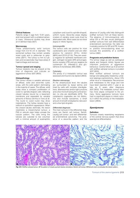
Fig. 3.14 Wolffian adnexal tumour. A The pattern of closely packed tubules simulates a Sertoli cell tumour. B Reticulin stain accentuates the tubular pattern.<br />
B<br />
Tumours of the uterine ligaments 213


