

Invasive breast carcinoma - IARC
Invasive breast carcinoma - IARC
Invasive breast carcinoma - IARC

Create successful ePaper yourself
Turn your PDF publications into a flip-book with our unique Google optimized e-Paper software.
A<br />
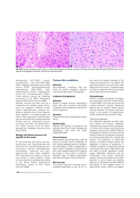
Fig. 2.124 Luteoma of pregnancy. A The tumour is composed of polygonal eosinophilic cells that form follicle-like spaces filled with pale fluid. B The tumour is composed<br />
of large polygonal eosinophilic cells that are mitotically actiive.<br />
B<br />
f i b ro s a rcoma {1517,1867}, leiomyo<br />
m y o s a rcoma {917,1416,1895,1983,<br />
2037}, malignant peripheral nerve sheath<br />
tumour {2797}, lymphangiosarc o m a ,<br />
a n g i o s a rcoma {2021,2064}, rhabd<br />
o m y o s a rcoma {2018}, osteosarc o m a<br />
{1215} and chondro s a rcoma {2851}.<br />
These tumours should be classified<br />
a c c o rding to the WHO Histological<br />
Typing of Soft Tissue Tumours {3086}.<br />
Similarly, tumours may also arise as a<br />
component of a complex ovarian tumour<br />
such as malignant müllerian mixed<br />
t u m o u r, adenosarcoma, immature teratoma<br />
or dermoid cyst or from heterologous<br />
elements in a Sertoli-Leydig cell<br />
tumour. Rare sarcomas of various types<br />
may be associated with surface epithelial<br />
s t romal tumours, particularly sero u s ,<br />
mucinous and clear cell adeno<strong>carcinoma</strong>.<br />
These tumours must be distinguished<br />
from metastatic sarcoma to the<br />
ovary {3222}.<br />
Benign soft tissue tumours not<br />
specific to the ovary<br />
Of the remaining soft tissue tumours,<br />
leiomyomas and haemangiomas are<br />
most common. Occasional benign neural<br />
tumours, lipomas, lymphangiomas,<br />
c h o n d romas, osteomas and gang<br />
l i o n e u romas have been re p o rt e d .<br />
Their appearance is similar to soft tissue<br />
tumours in other locations. These<br />
tumours should be classified accord i n g<br />
to the World Health Organization<br />
Histological Typing of Soft Tissue<br />
Tumours {3086}.<br />
Tumour-like conditions<br />
Definition<br />
Non-neoplastic conditions that can<br />
mimic an ovarian neoplasm clinically,<br />
macroscopically and/or histologically.<br />
Luteoma of pregnancy<br />
Definition<br />
Single or multiple nodules composed of<br />
lutein cells with abundant eosinophilic<br />
cytoplasm that are detected at the end of<br />
a term pregnancy.<br />
Synonym<br />
Nodular theca-lutein hyperplasia of pregnancy.<br />
Epidemiology<br />
Patients with luteoma of pregnancy are<br />
typically in their third or fourth decade and<br />
m u l t i p a rous, and 80% are Black<br />
{ 2 0 5 6 , 2 3 6 4 , 2 7 8 8 } .<br />
Clinical features<br />
Most patients are asymptomatic, and the<br />
tumour is usually found incidentally at<br />
t e rm during caesarean section or postpartum<br />
tubal ligation {2788}. Exceptionally, a<br />
pelvic mass is palpable or obstructs the<br />
b i rth canal. Approximately 25% of patients<br />
a re hirsute or show signs of virilization.<br />
Elevated levels of plasma testostero n e<br />
and other androgens may be observed.<br />
Macrosocopy<br />
The tumours vary from not being macroscopically<br />
detectable to over 20 cm. In<br />
one series the medium diameter of the<br />
tumour was between 6-7 cm {2056}. The<br />
sectioned surface is circumscribed, solid,<br />
fleshy and red to brown. In appro x i m a t e l y<br />
one-half of cases the lesions are multiple<br />
and at least one-third are bilateral.<br />
Histopathology<br />
There is a diffuse proliferation of polygonal,<br />
eosinophilic cells that contain little or<br />
no lipid {2364}. The nuclei are round and<br />
contain prominent nucleoli. Follicle-like<br />
spaces may be present. Mitotic figures<br />
may be frequent. The tumour cells were<br />
found to be positive for alpha-inhibin,<br />
CD99, cytokeratin and vimentin {2242}.<br />
Differential diagnosis<br />
The diff e rential diagnosis includes lipidpoor<br />
steroid cell tumours, metastatic<br />
melanoma and corpus luteum of pre g n a n-<br />
c y. Steroid cell tumours occurring during<br />
p regnancy may present a difficult diff e re n-<br />
tial diagnosis; however, the typical clinical<br />
setting of luteoma of pregnancy would be<br />
an unusual presentation for a steroid cell<br />
t u m o u r. The presence of follicle-like<br />
spaces or multiple nodules favours the<br />
diagnosis of luteoma of pre g n a n c y. In<br />
contrast to luteoma of pre g n a n c y, stero i d<br />
cell tumours that have a high mitotic rate<br />
a re likely to exhibit significant nuclear<br />
atypia. Metastatic melanoma may be<br />
multinodular and contain follicle-like<br />
spaces; however, the presence of melanin<br />
pigment in some cases and positive<br />
stains for S-100 protein and often HMB-45<br />
and Melan A and negative stains for<br />
alpha-inhibin would confirm the diagno-<br />
188 Tumours of the ovary and peritoneum


