

Invasive breast carcinoma - IARC
Invasive breast carcinoma - IARC
Invasive breast carcinoma - IARC

Create successful ePaper yourself
Turn your PDF publications into a flip-book with our unique Google optimized e-Paper software.
used. Inflammatory conditions in the<br />
b reast may mimic MALT lymphoma.<br />
Prognosis and predictive factors<br />
P r i m a ry <strong>breast</strong> lymphomas behave in a<br />
way similar to lymphomas of corre s p o n-<br />
ding type and stage in other sites.<br />
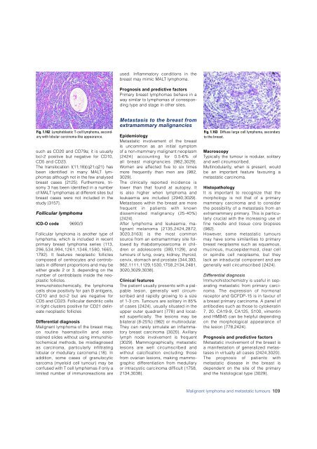
Fig. 1.162 Lymphoblastic T-cell lymphoma, secondary<br />
with lobular <strong>carcinoma</strong>-like appearance.<br />
such as CD20 and CD79a; it is usually<br />
bcl-2 positive but negative for CD10,<br />
CD5 and CD23.<br />
The translocation t(11;18)(q21;q21) has<br />
been identified in many MALT lymphomas<br />
although not in the few analysed<br />
<strong>breast</strong> cases {2125}. Furthermore, trisomy<br />
3 has been identified in a number<br />
of MALT lymphomas at different sites but<br />
<strong>breast</strong> cases were not included in the<br />
study {3157}.<br />
Follicular lymphoma<br />
ICD-O code 9 6 9 0 / 3<br />
Follicular lymphoma is another type of<br />
lymphoma, which is included in re c e n t<br />
p r i m a ry <strong>breast</strong> lymphoma series {113,<br />
2 9 6 , 5 3 4 , 9 9 4 , 1 2 6 1 , 1 3 4 6 , 1 5 8 0 , 1 6 6 5 ,<br />
1792}. It features neoplastic follicles<br />
composed of centrocytes and centro b-<br />
lasts in diff e rent pro p o rtions and may be<br />
either grade 2 or 3, depending on the<br />
number of centroblasts inside the neoplastic<br />
follicles.<br />
I m m u n o h i s t o c h e m i c a l l y, the lymphoma<br />
cells show positivity for pan B antigens,<br />
CD10 and bcl-2 but are negative for<br />
CD5 and CD23. Follicular dendritic cells<br />
in tight clusters positive for CD21 delineate<br />
neoplastic follicles<br />
Differential diagnosis<br />
Malignant lymphoma of the <strong>breast</strong> may,<br />
on routine haematoxilin and eosin<br />
stained slides without using immunohistochemical<br />
methods, be misdiagnosed<br />
as <strong>carcinoma</strong>, particularly infiltrating<br />
lobular or medullary <strong>carcinoma</strong> {18}. In<br />
addition, some cases of granulocytic<br />
s a rcoma (myeloid cell tumour) may be<br />
confused with T cell lymphomas if only a<br />
limited number of immunoreactions are<br />
Metastasis to the <strong>breast</strong> from<br />
extramammary malignancies<br />
E p i d e m i o l o g y<br />
Metastatic involvement of the <strong>breast</strong><br />
is uncommon as an initial symptom<br />
of a non-mammary malignant neoplasm<br />
{2424} accounting for 0.5-6% of<br />
all b reast malignancies {982,3029}.<br />
Women are affected five to six times<br />
m o re frequently than men are {982,<br />
3 0 2 9 } .<br />
The clinically re p o rted incidence is<br />
lower than that found at autopsy. It<br />
is also higher when lymphoma and<br />
leukaemia are included {2940,3029}.<br />
Metastases within the <strong>breast</strong> are more<br />
f requent in patients with known<br />
disseminated malignancy (25-40%)<br />
{2424}.<br />
After lymphoma and leukaemia, malignant<br />
melanoma {2135,2424,2872,<br />
3020,3163} is the most common<br />
s o u rce from an extramammary site followed<br />
by rhabdomyosarcoma in child<br />
ren or adolescents {393,1129}, and<br />
tumours of lung, ovary, kidney, thyro i d ,<br />
cervix, stomach and prostate {344,393,<br />
9 8 2 , 1 1 1 1 , 1 1 2 9 , 1 5 3 0 , 1 7 5 8 , 2 1 3 4 , 2 4 8 1 ,<br />
3 0 2 0 , 3 0 2 9 , 3 0 3 8 } .<br />
Clinical features<br />
The patient usually presents with a palpable<br />
lesion, generally well circ u m-<br />
scribed and rapidly growing to a size<br />
of 1-3 cm. Tumours are solitary in 85%<br />
of cases {2424}, usually situated in the<br />
upper outer quadrant {778} and located<br />
superf i c i a l l y. The lesions may be<br />
bilateral (8-25%) {982} or multinodular.<br />
They can rarely simulate an inflammat<br />
o ry <strong>breast</strong> <strong>carcinoma</strong> {3020}. Axillary<br />
lymph node involvement is fre q u e n t<br />
{3029}. Mammographically, metastatic<br />
lesions are well circumscribed and<br />
without calcification excluding those<br />
f rom ovarian lesions, making mammographic<br />
diff e rentiation from medullary<br />
or intracystic <strong>carcinoma</strong> difficult {1758,<br />
2 1 3 4 , 3 0 3 8 } .<br />
Fig. 1.163 Diffuse large cell lymphoma, secondary<br />
to the <strong>breast</strong>.<br />
M a c r o s c o p y<br />
Typically the tumour is nodular, solitary<br />
and well circ u m s c r i b e d .<br />
M u l t i n o d u l a r i t y, when is present, would<br />
be an important feature favouring a<br />
metastatic carc i n o m a .<br />
H i s t o p a t h o l o g y<br />
It is important to recognize that the<br />
morphology is not that of a primary<br />
m a m m a ry <strong>carcinoma</strong> and to consider<br />
the possibility of a metastasis from an<br />
e x t r a m a m m a ry primary. This is part i c u-<br />
larly crucial with the increasing use of<br />
fine needle and tissue core biopsies<br />
{982}.<br />
H o w e v e r, some metastatic tumours<br />
may have some similarities to primary<br />
b reast neoplasms such as squamous,<br />
mucinous, mucoepidermoid, clear cell<br />
or spindle cell neoplasms, but they<br />
lack an intraductal component and are<br />
generally well circumscribed {2424}.<br />
Differential diagnosis<br />
I m m u n o h i s t o c h e m i s t ry is useful in separating<br />
metastatic from primary carc i-<br />
noma. The expression of horm o n a l<br />
receptor and GCFDP-15 is in favour of<br />
a <strong>breast</strong> primary <strong>carcinoma</strong>. A panel of<br />
antibodies such as those to cytokeratin<br />
7, 20, CA19-9, CA125, S100, vimentin<br />
and HMB45 can be helpful depending<br />
on the morphological appearance of<br />
the lesion {778,2424}.<br />
Prognosis and predictive factors<br />
Metastatic involvement of the <strong>breast</strong> is<br />
a manifestation of generalized metastases<br />
in virtually all cases {2424,3020}.<br />
The prognosis of patients with<br />
metastatic disease in the <strong>breast</strong> is<br />
dependent on the site of the primary<br />
and the histological type {3029} .<br />
Malignant lymphoma and metastatic tumours 109


