

Invasive breast carcinoma - IARC
Invasive breast carcinoma - IARC
Invasive breast carcinoma - IARC

Create successful ePaper yourself
Turn your PDF publications into a flip-book with our unique Google optimized e-Paper software.
Melanocytic tumours<br />
E.J. Wilkinson<br />
M.R. Teixeira<br />
Malignant melanomas account for 2-10%<br />
of vulvar malignancies {2316} and occur<br />
predominantly in elderly White women. A<br />
variety of naevi that must be distinguished<br />
from melanoma also occur in the<br />
vulva.<br />
ICD-O codes<br />
Malignant melanoma 8720/3<br />
Congenital melanocytic naevus 8761/0<br />
Acquired melanocytic naevus 8720/0<br />
Blue naevus 8780/0<br />
Atypical melanocytic naevus<br />
of the genital type 8720/0<br />
Dysplastic melanocytic naevus 8727/0<br />
Malignant melanoma<br />
Definition<br />
A malignant tumour of melanocytic origin.<br />
Clinical features<br />
Signs and symptoms<br />
Symptoms include vulvar bleeding, pruritus<br />
and dysuria. Although vulvar malignant<br />
melanoma usually presents as a<br />
pigmented mass, 27% are non-pigmented<br />
{2320}. Satellite cutaneous nodules<br />
occur in 20% of cases {2320}. Melanoma<br />
may arise in a prior benign or atypical<br />
appearing melanocytic lesion. {1912,<br />
3151}. The majority present as a nodule<br />
or polypoid mass. Approximately 5% are<br />
ulcerated {2320}. They occur with nearly<br />
equal frequency in the labia majora, labia<br />
minora or clitoris.<br />
Imaging<br />
Radiological, magnetic resonance imaging<br />
and/or radiolabelled isotope scan<br />
studies may be used to assess tumour<br />
that is present outside the vulva.<br />
Histopathology<br />
Three histological types of melanoma are<br />
identified: superficial spreading, nodular<br />
and mucosal/acral lentiginous {216,<br />
1355,2261,2864}. Approximately 25% of<br />
the cases are unclassifiable {2320}.<br />
Melanomas may be composed of epithelioid,<br />
spindle, dendritic, nevoid or mixed<br />
cell types. The epithelioid cells contain<br />
abundant eosinophilic cytoplasm, large<br />
nuclei and prominent nucleoli. The dendritic<br />
cells have tapering cytoplasmic<br />
extensions resembling nerve cells and<br />
show moderate nuclear pleomorphism.<br />
Spindle-shaped cells have smaller, oval<br />
nuclei and may be arranged in sheets or<br />
bundles. Certain cell types may predominate<br />
within a given tumour. The amount<br />
of melanin within the tumour cells is highly<br />
variable, and cells may contain no<br />
melanin.<br />
Both mucosal/acral lentiginous and<br />
superficial spreading melanomas can be<br />
e n t i rely intraepithelial. When invasive,<br />
both histological types have vertical and<br />
radial growth phases, the vertical growth<br />
component re p resenting the invasive<br />
focus of tumour. Nodular melanomas display<br />
predominately a vertical gro w t h<br />
phase. Atypical melanocytes characteristic<br />
of melanoma in situ usually can be<br />
identified within the epithelium adjacent<br />
to mucosal/acral lentiginous and superficial<br />
spreading melanomas.<br />
Superficial spreading melanomas have<br />
malignant melanocytic cells within the<br />
area of invasion that are typically large<br />
with relatively uniform nuclei and prominent<br />
nucleoli, similar to the adjacent<br />
intraepithelial melanoma. The intraepithelial<br />
component is considered to be<br />
the radial growth portion of the tumour.<br />
Nodular melanomas may have a small<br />
neoplastic intraepithelial component<br />
adjacent to the invasive tumour and are<br />
generally not considered to have a significant<br />
radial growth phase. The cells of<br />
nodular melanomas may be epithelioid or<br />
spindle-shaped. These tumours are typically<br />
deeply invasive.<br />
Mucosal/acral lentiginous melanomas<br />
a re most common within the vulvar<br />
vestibule, including the clitoris. They are<br />
characterized by spindle-shaped neoplastic<br />
melanocytes within the junctional<br />
zone involving the adjacent superficial<br />
stroma in a diffuse pattern. The spindleshaped<br />
cells are relatively uniform, lacking<br />
significant nuclear pleomorphism.<br />
Within the stroma the tumour is usually<br />
associated with a desmoplastic<br />
response.<br />
There is some variation in the reported<br />
frequency of melanoma types involving<br />
the vulva; however, in a large series of<br />
198 cases mucosal/acral lentiginous<br />
melanoma comprised 52% of the cases,<br />
nodular melanoma 20% and superficial<br />
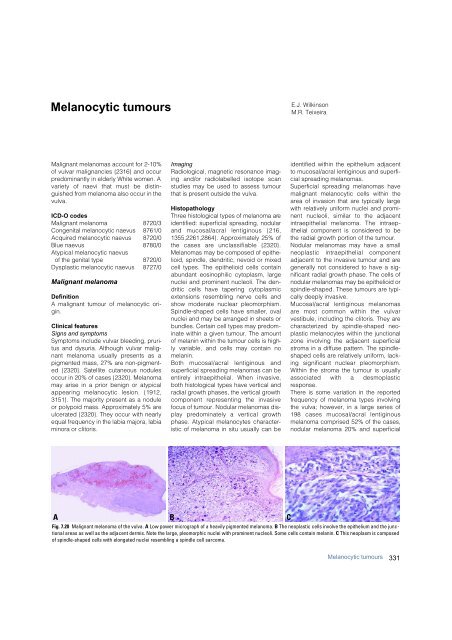
A<br />
B<br />
Fig. 7.20 Malignant melanoma of the vulva. A Low power micrograph of a heavily pigmented melanoma. B The neoplastic cells involve the epithelium and the junctional<br />
areas as well as the adjacent dermis. Note the large, pleomorphic nuclei with prominent nucleoli. Some cells contain melanin. C This neoplasm is composed<br />
of spindle-shaped cells with elongated nuclei resembling a spindle cell sarcoma.<br />
C<br />
Melanocytic tumours<br />
331


