

Invasive breast carcinoma - IARC
Invasive breast carcinoma - IARC
Invasive breast carcinoma - IARC

Create successful ePaper yourself
Turn your PDF publications into a flip-book with our unique Google optimized e-Paper software.
administration or to endogenous hyperoestrinism<br />
{2276,2648,2805}. Endometrial<br />
hyperplasia and atypical hyperplasia<br />
have similar clinical associations.<br />
Imaging<br />
Transvaginal ultrasound (US) is the imaging<br />
technique of choice for the assessment<br />
of the endometrium in symptomatic<br />
patients, e.g. in cases of postmenopausal<br />
bleeding {133}. In postmenopausal<br />
women without hormonal replacement an<br />
endometrial thickness of 5 mm is re g a rded<br />
as the upper normal limit {133,2650}.<br />
The presence of endometrial thickening<br />
on ultrasound or cross sectional imaging<br />
is, however, a nonspecific finding. It may<br />
be due to endometrial hyperplasia,<br />
polyps or <strong>carcinoma</strong>. The final diagnosis<br />
usually needs to be determined by<br />
endometrial sampling {133}.<br />
Whereas currently magnetic resonance<br />
imaging (MRI) has no established role in<br />
screening for endometrial pathology, it is<br />
regarded as the best imaging technique<br />
for preoperative staging of endometrial<br />
<strong>carcinoma</strong> proven by endometrial sampling.<br />
MRI was shown to be superior to<br />
computed tomography (CT) in this<br />
regard {1135}. It is especially useful for<br />
patients with suspected advanced disease,<br />
for those with associated uterine<br />
pathology, such as leiomyomas, and for<br />
those with histological subtypes that signify<br />
a worse prognosis {916,1136}.<br />
Macroscopy<br />
Endometrial <strong>carcinoma</strong> usually arises in<br />
the uterine corpus, but some cases originate<br />
in the lower uterine segment, and<br />
recent studies suggest that the latter may<br />
have diff e rent clinical and histological feat<br />
u res {1323,3067}. Regardless of the histological<br />
type, the macroscopic appearance<br />
of endometrial <strong>carcinoma</strong> is generally<br />
that of a single dominant mass, usually<br />
occurring in an enlarged uterus, although<br />
occasionally the uterus is small or the<br />
tumour presents as a diffuse thickening of<br />
most of the endometrial surface, part i c u-<br />
larly in the serous type. Endometrial carc i-<br />
noma is seen more frequently on the posterior<br />
than on the anterior wall {2691}.<br />
The typical <strong>carcinoma</strong> is exophytic and<br />
has a shaggy, frequently ulcerated surface<br />
beneath which a soft or firm white<br />
tumour may extend shallowly or deeply<br />
into the underlying myometrium. In<br />
advanced cases the tumour may penetrate<br />
the serosa or extend into the cervix.<br />
An estimate of the extent of tumour may<br />
be requested preoperatively or operatively<br />
in order to determine the extent of the<br />
surgical pro c e d u re to be perf o rmed {594}.<br />
In occasional cases no tumour may be<br />
visible macro s c o p i c a l l y, with carc i n o m a<br />
identified only at histological examination.<br />
Tumour spread and staging<br />
The staging of uterine tumours is by the<br />
TNM/FIGO classification {51,2976}.<br />
Endometrioid adeno<strong>carcinoma</strong><br />
Definition<br />
A primary endometrial adeno<strong>carcinoma</strong><br />
containing glands resembling those of<br />
the normal endometrium.<br />
Histopathology<br />
All but a few rare endometrial <strong>carcinoma</strong>s<br />
are adeno<strong>carcinoma</strong>s, and the most<br />
common of these is the endometrioid<br />
type {2691}. Endometrioid adeno<strong>carcinoma</strong><br />
represents a spectrum of histological<br />
differentiation from a very well differentiated<br />
<strong>carcinoma</strong> difficult to distinguish<br />
f rom atypical complex hyperplasia to<br />
minimally differentiated tumours that can<br />
be confused not only with undifferentiated<br />
<strong>carcinoma</strong> but with various sarcomas<br />
as well. A highly characteristic feature of<br />
endometrioid adeno<strong>carcinoma</strong> is the<br />
presence of at least some glandular or<br />
villoglandular structures lined by simple<br />
to pseudostratified columnar cells that<br />
have their long axes arranged perpendicular<br />
to the basement membrane with at<br />
least somewhat elongated nuclei that are<br />
also polarized in the same direction. As<br />
the glandular differentiation decreases<br />
and is replaced by solid nests and<br />
sheets of cells, the tumour is classified as<br />
less well differentiated (higher grade).<br />
Deep myometrial invasion and lymph<br />
node metastases are both more frequent<br />
in higher grade <strong>carcinoma</strong>s, and survival<br />
rates are correspondingly lower {574,<br />
1359}. It should be noted that:<br />
(1). Only those cells which are considered<br />
to be of glandular type are considered<br />
in the grading schema, so that solid<br />
nests of cells showing squamous or<br />
morular differentiation do not increase<br />
the tumour grade.<br />
(2). Bizarre nuclear atypia should raise the<br />
grade by one, e.g. from 1 to 2 or 2 to 3.<br />
(3). It should be emphasized that the<br />
presence of bizarre nuclei occurring in<br />
even a predominantly glandular tumour<br />
may indicate serous or clear cell rather<br />
than endometrioid differentiation {2691}.<br />
The distinction of very well differentiated<br />
A<br />
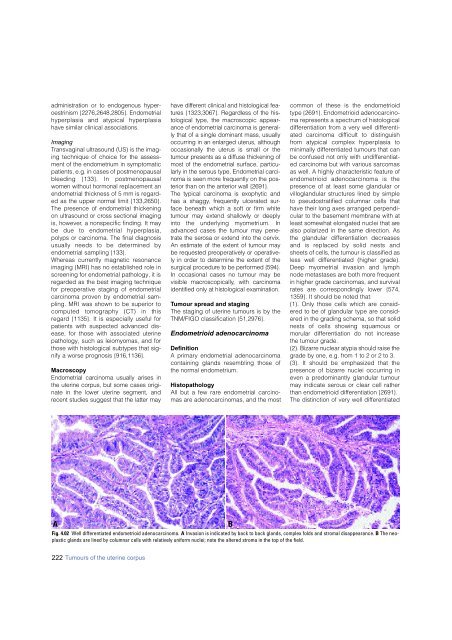
Fig. 4.02 Well differentiated endometrioid adeno<strong>carcinoma</strong>. A Invasion is indicated by back to back glands, complex folds and stromal disappearance. B The neoplastic<br />
glands are lined by columnar cells with relatively uniform nuclei; note the altered stroma in the top of the field.<br />
B<br />
222 Tumours of the uterine corpus


