

Invasive breast carcinoma - IARC
Invasive breast carcinoma - IARC
Invasive breast carcinoma - IARC

Create successful ePaper yourself
Turn your PDF publications into a flip-book with our unique Google optimized e-Paper software.
Table 6.01<br />
Terminology of premalignant vaginal squamous epithelial lesions.<br />
Classification<br />
Synonyms<br />
Vaginal intraepithelial Mild dysplasia Low grade VAIN<br />
neoplasia, grade 1<br />
Vaginal intraepithelial Moderate dysplasia High grade VAIN<br />
neoplasia, grade 2<br />
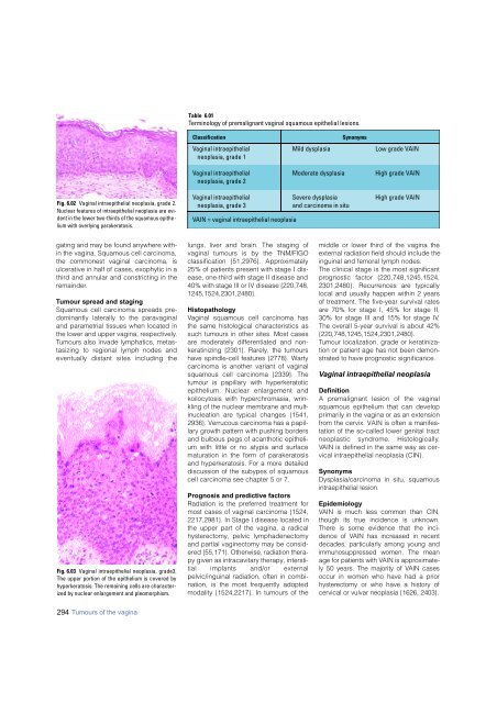
Fig. 6.02 Vaginal intraepithelial neoplasia, grade 2.<br />
Nuclear features of intraepithelial neoplasia are evident<br />
in the lower two-thirds of the squamous epithelium<br />
with overlying parakeratosis.<br />
Vaginal intraepithelial Severe dysplasia High grade VAIN<br />
neoplasia, grade 3<br />
and <strong>carcinoma</strong> in situ<br />
VAIN = vaginal intraepithelial neoplasia<br />
gating and may be found anywhere within<br />
the vagina. Squamous cell <strong>carcinoma</strong>,<br />
the commonest vaginal <strong>carcinoma</strong>, is<br />
ulcerative in half of cases, exophytic in a<br />
third and annular and constricting in the<br />
remainder.<br />
Tumour spread and staging<br />
Squamous cell <strong>carcinoma</strong> spreads predominantly<br />
laterally to the paravaginal<br />
and parametrial tissues when located in<br />
the lower and upper vagina, respectively.<br />
Tumours also invade lymphatics, metastasizing<br />
to regional lymph nodes and<br />
eventually distant sites including the<br />
Fig. 6.03 Vaginal intraepithelial neoplasia, grade3.<br />
The upper portion of the epithelium is covered by<br />
hyperkeratosis. The remaining cells are characterized<br />
by nuclear enlargement and pleomorphism.<br />
lungs, liver and brain. The staging of<br />
vaginal tumours is by the TNM/FIGO<br />
classification {51,2976}. Appro x i m a t e l y<br />
25% of patients present with stage I disease,<br />
one-third with stage II disease and<br />
40% with stage III or IV disease {220,748,<br />
1245,1524,2301,2480}.<br />
Histopathology<br />
Vaginal squamous cell <strong>carcinoma</strong> has<br />
the same histological characteristics as<br />
such tumours in other sites. Most cases<br />
are moderately differentiated and nonkeratinizing<br />
{2301}. Rarely, the tumours<br />
have spindle-cell features {2778}. Warty<br />
<strong>carcinoma</strong> is another variant of vaginal<br />
squamous cell <strong>carcinoma</strong> {2339}. The<br />
tumour is papillary with hyperkeratotic<br />
epithelium. Nuclear enlargement and<br />
koilocytosis with hyperchromasia, wrinkling<br />
of the nuclear membrane and multinucleation<br />
are typical changes {1541,<br />
2936}. Verrucous <strong>carcinoma</strong> has a papillary<br />
growth pattern with pushing borders<br />
and bulbous pegs of acanthotic epithelium<br />
with little or no atypia and surface<br />
maturation in the form of parakeratosis<br />
and hyperkeratosis. For a more detailed<br />
discussion of the subypes of squamous<br />
cell <strong>carcinoma</strong> see chapter 5 or 7.<br />
Prognosis and predictive factors<br />
Radiation is the preferred treatment for<br />
most cases of vaginal <strong>carcinoma</strong> {1524,<br />
2217,2981}. In Stage I disease located in<br />
the upper part of the vagina, a radical<br />
hysterectomy, pelvic lymphadenectomy<br />
and partial vaginectomy may be considered<br />
{55,171}. Otherwise, radiation therapy<br />
given as intracavitary therapy, interstitial<br />
implants and/or extern a l<br />
pelvic/inguinal radiation, often in combination,<br />
is the most frequently adopted<br />
modality {1524,2217}. In tumours of the<br />
middle or lower third of the vagina the<br />
external radiation field should include the<br />
inguinal and femoral lymph nodes.<br />
The clinical stage is the most significant<br />
p rognostic factor {220,748,1245,1524,<br />
2301,2480}. Recurrences are typically<br />
local and usually happen within 2 years<br />
of treatment. The five-year survival rates<br />
are 70% for stage I, 45% for stage II,<br />
30% for stage III and 15% for stage IV.<br />
The overall 5-year survival is about 42%<br />
{220,748,1245,1524,2301,2480}.<br />
Tumour localization, grade or keratinization<br />
or patient age has not been demonstrated<br />
to have prognostic significance.<br />
Vaginal intraepithelial neoplasia<br />
Definition<br />
A premalignant lesion of the vaginal<br />
squamous epithelium that can develop<br />
primarily in the vagina or as an extension<br />
from the cervix. VAIN is often a manifestation<br />
of the so-called lower genital tract<br />
neoplastic syndrome. Histologically,<br />
VAIN is defined in the same way as cervical<br />
intraepithelial neoplasia (CIN).<br />
Synonyms<br />
Dysplasia/<strong>carcinoma</strong> in situ, squamous<br />
intraepithelial lesion.<br />
Epidemiology<br />
VAIN is much less common than CIN,<br />
though its true incidence is unknown.<br />
T h e re is some evidence that the incidence<br />
of VAIN has increased in re c e n t<br />
decades, particularly among young and<br />
i m m u n o s u p p ressed women. The mean<br />
age for patients with VAIN is appro x i m a t e-<br />
ly 50 years. The majority of VAIN cases<br />
occur in women who have had a prior<br />
h y s t e rectomy or who have a history of<br />
cervical or vulvar neoplasia {1626, 2403}.<br />
294 Tumours of the vagina


