

Invasive breast carcinoma - IARC
Invasive breast carcinoma - IARC
Invasive breast carcinoma - IARC

Create successful ePaper yourself
Turn your PDF publications into a flip-book with our unique Google optimized e-Paper software.
cases, anisokaryosis has no prognostic<br />
implication The behaviour of dysgerminoma<br />
with trophoblastic differentiation is<br />
identical to the usual type, but with the<br />
advantage of having β-hCG as a serum<br />
marker.<br />
Yolk sac tumour<br />
Definition<br />
Yolk sac tumours are morphologically<br />
h e t e rogeneous, primitive teratoid neoplasms<br />
differentiating into multiple endodermal<br />
structures, ranging from the primitive<br />
gut to its derivatives of extraembryonal<br />
(secondary yolk sac vesicle) and<br />
embryonal somatic type, e.g. intestine,<br />
liver {2035}. These neoplasms have<br />
many epithelial patterns and are typically<br />
immunoreactive for alpha-fetoprotein.<br />
Synonym and historical annotation<br />
Since the secondary yolk sac component<br />
represents only one of its many lines of<br />
differentiation, the current nomenclature<br />
is clearly restrictive. Perhaps the term<br />
“endodermal primitive tumours” would<br />
be more accurate in defining all the possible<br />
lines of differentiation, both epithelial<br />
and mesenchymal, that occur in<br />
these neoplasms.<br />
The term “endodermal sinus tumour”,<br />
although still in use, is misleading, since<br />
the endodermal sinus is neither a structure<br />
present in human embryogenesis<br />
{1463} nor is it a constant feature of these<br />
neoplasms, as it only occurs in a minority<br />
of cases {1537}.<br />
Macroscopy<br />
These tumours are usually well encapsulated<br />
with an average diameter of 15 cm<br />
{1537}. The sectioned tumour surface is<br />
soft and grey-yellow with frequent areas<br />
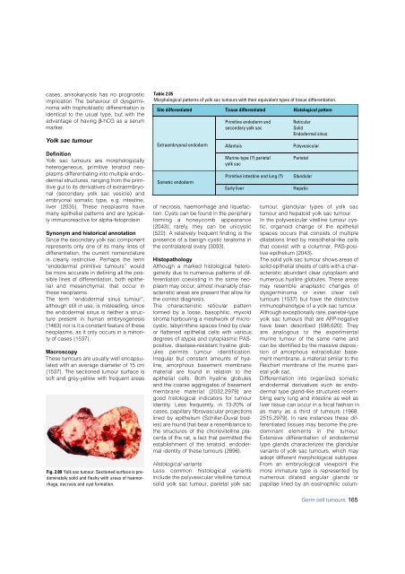
Fig. 2.89 Yolk sac tumour. Sectioned surface is predominately<br />
solid and fleshy with areas of haemorrhage,<br />
necrosis and cyst formation.<br />
Table 2.05<br />
Morphological patterns of yolk sac tumours with their equivalent types of tissue differentiation.<br />
Site differentiated Tissue differentiated Histological pattern<br />
Extraembryonal endoderm<br />
Somatic endoderm<br />
Primitive endoderm and<br />
secondary yolk sac<br />
Allantois<br />
Murine-type (?) parietal<br />
yolk sac<br />
of necrosis, haemorrhage and liquefaction.<br />
Cysts can be found in the periphery<br />
f o rming a honeycomb appearance<br />
{2043}; rare l y, they can be unicystic<br />
{522}. A relatively frequent finding is the<br />
presence of a benign cystic teratoma in<br />
the contralateral ovary {3033}.<br />
Histopathology<br />
Although a marked histological heterogeneity<br />
due to numerous patterns of differentiation<br />
coexisting in the same neoplasm<br />
may occur, almost invariably characteristic<br />
areas are present that allow for<br />
the correct diagnosis.<br />
The characteristic reticular pattern<br />
formed by a loose, basophilic, myxoid<br />
stroma harbouring a meshwork of microcystic,<br />
labyrinthine spaces lined by clear<br />
or flattened epithelial cells with various<br />
degrees of atypia and cytoplasmic PASpositive,<br />
diastase-resistant hyaline globules<br />
permits tumour identification.<br />
Irregular but constant amounts of hyaline,<br />
amorphous basement membrane<br />
material are found in relation to the<br />
epithelial cells. Both hyaline globules<br />
and the coarse aggregates of basement<br />
membrane material {2032,2979} are<br />
good histological indicators for tumour<br />
identity. Less frequently, in 13-20% of<br />
cases, papillary fibrovascular projections<br />
lined by epithelium (Schiller-Duval bodies)<br />
are found that bear a resemblance to<br />
the structures of the choriovitelline placenta<br />
of the rat, a fact that permitted the<br />
establishment of the teratoid, endodermal<br />
identity of these tumours {2896}.<br />
Histological variants<br />
Less common histological variants<br />
include the polyvesicular vitelline tumour,<br />
solid yolk sac tumour, parietal yolk sac<br />
Primitive intestine and lung (?)<br />
Early liver<br />
Reticular<br />
Solid<br />
Endodermal sinus<br />
Polyvesicular<br />
Parietal<br />
Glandular<br />
Hepatic<br />
t u m o u r, glandular types of yolk sac<br />
tumour and hepatoid yolk sac tumour.<br />
In the polyvesicular vitelline tumour cystic,<br />
organoid change of the epithelial<br />
spaces occurs that consists of multiple<br />
dilatations lined by mesothelial-like cells<br />
that coexist with a columnar, PAS-positive<br />
epithelium {2043}.<br />
The solid yolk sac tumour shows areas of<br />
solid epithelial sheets of cells with a characteristic<br />
abundant clear cytoplasm and<br />
numerous hyaline globules. These areas<br />
may resemble anaplastic changes of<br />
d y s g e rminoma or even clear cell<br />
tumours {1537} but have the distinctive<br />
immunophenotype of a yolk sac tumour.<br />
Although exceptionally rare, parietal-type<br />
yolk sac tumours that are AFP-negative<br />
have been described {598,620}. They<br />
a re analogous to the experimental<br />
murine tumour of the same name and<br />
can be identified by the massive deposition<br />
of amorphous extracellular basement<br />
membrane, a material similar to the<br />
Reichert membrane of the murine parietal<br />
yolk sac.<br />
D i ff e rentiation into organized somatic<br />
endodermal derivatives such as endodermal<br />
type gland-like structures resembling<br />
early lung and intestine as well as<br />
liver tissue can occur in a focal fashion in<br />
as many as a third of tumours {1968,<br />
2515,2979}. In rare instances these differentiated<br />
tissues may become the predominant<br />
elements in the tumour.<br />
Extensive differentiation of endodermal<br />
type glands characterizes the glandular<br />
variants of yolk sac tumours, which may<br />
adopt different morphological subtypes.<br />
F rom an embryological viewpoint the<br />
more immature type is represented by<br />
n u m e rous dilated angular glands or<br />
papillae lined by an eosinophilic colum-<br />
Germ cell tumours 165


