

Invasive breast carcinoma - IARC
Invasive breast carcinoma - IARC
Invasive breast carcinoma - IARC

You also want an ePaper? Increase the reach of your titles
YUMPU automatically turns print PDFs into web optimized ePapers that Google loves.
have a strong association with high grade<br />
CIN lesions {587,1088,1699, 1878,3127}.<br />
CIN re p resents the preinvasive counterp<br />
a rt of invasive cervical squamous cell<br />
c a rcinoma, and there is now abundant<br />
evidence for its malignant potential.<br />
H o w e v e r, there is no inevitability about<br />
neoplastic pro g ression; such lesions may<br />
re g ress, remain phenotypically stable or<br />
p ro g ress {2137}.<br />
Histopathology<br />
C o n v e n t i o n a l l y, these are subjectively<br />
divided into three grades: CIN 1, 2 and 3,<br />
though the histological features re p re s e n t<br />
a diagnostic continuum. Incre a s i n g - l y,<br />
t h e re is a tendency to use a two-tiere d<br />
classification of low and high grade CIN<br />
that equates to CIN 1 and CIN 2 and 3<br />
re s p e c t i v e l y. These precursors may also<br />
be re f e r red to as low and high grade<br />
squamous intraepithelial lesion (SIL)<br />
{2177}. Because of the inherent diff i c u l t y<br />
in distinguishing pure HPV infection fro m<br />
unequivocal CIN 1 in flat, non-condylomatous<br />
epithelium (sometimes confusingly<br />
re f e r red to as flat condyloma), HPV infection<br />
alone is included in the low grade SIL<br />
c a t e g o ry, a terminology that has been<br />
m o re widely accepted by cytopathologists<br />
{1612}. The relationship of the varying<br />
terminology is shown in Table 5.1.<br />
A<br />
B<br />
Fig. 5.14 A Cervical intraepithelial neoplasia (CIN 1).<br />
The upper two-thirds of the epithelium show maturation<br />
and focal koilocytosis. There is mild atypia throughout.<br />
B CIN 2. Nuclear abnormalities are more striking than in<br />
CIN 1, and mitoses are seen (centre). The upper third of<br />
the epithelium shows maturation.<br />
Cervical intraepithelial neoplasia 1<br />
Maturation is present in the upper twothirds<br />
of the epithelium, and the superficial<br />
cells contain variable but usually<br />
mild atypia, which may include viral cytopathic<br />
effect (koilocytosis). Nuclear<br />
abnormalities are present throughout but<br />
are slight. Mitotic figures are present in<br />
the basal third and are not numerous.<br />
Abnormal forms are rare.<br />
Cervical intraepithelial neoplasia 2<br />
Maturation is present in the upper half of<br />
the epithelium, and nuclear atypia is conspicuous<br />
in both the upper and lower<br />
epithelial layers. Mitotic figures are generally<br />
confined to the basal two-thirds of the<br />
epithelium. Abnormal forms may be seen.<br />
Cervical intraepithelial neoplasia 3<br />
Maturation (including surface keratinization)<br />
may be absent or confined to the<br />
s u p e rficial third of the epithelium. Nuclear<br />
a b n o rmalities are marked thro u g h o u t<br />
most or all of the thickness of the epithelium.<br />
Mitotic figures may be numero u s<br />
and are found at all levels of the epithelium.<br />
Abnormal mitoses are fre q u e n t .<br />
Growth fraction<br />
HPVs, particularly high risk HPVs, are<br />
associated with alterations in the cell<br />
A<br />
B<br />
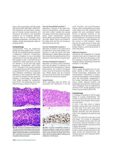
Fig. 5.15 A Cervical intraepithelial neoplasia 3.<br />
Squamous epithelium consists entirely of atypical<br />
basaloid cells. Note the moderate nuclear polymorphism,<br />
coarse chromatin and mitotic figures in the<br />
upper half of the epithelium. B Ki-67 staining shows<br />
proliferation in all cell layers.<br />
cycle. Therefore, cell cycle biomarkers<br />
may be useful in distinguishing non-diagnostic<br />
atypia from CIN. Expression of a<br />
generic cell cycle proliferation marker<br />
(Ki-67) is typically confined to the<br />
suprabasal cells of the lower third of the<br />
normal epithelium. The presence of Ki-67<br />
positive cells in the upper epithelial layers<br />
occurs in HPV infection, which<br />
induces cell cycle activity in these cells<br />
{1881,2356}. P16 ink4 , a cyclin-dependent<br />
kinase inhibitor, is a promising marker of<br />
CIN {1422,2527}.<br />
Differential diagnosis<br />
Transitional cell metaplasia is a benign<br />
condition that may be mistaken for high<br />
grade CIN. After the menopause immature<br />
squamous mucosa may exhibit histological<br />
features resembling transitional<br />
epithelium {1140,3085}.<br />
Related lesions<br />
CIN is usually associated with the cytopathic<br />
effects of HPV infection, which<br />
include koilocytosis, dyskeratosis and<br />
multinucleation. Koilocytosis is characterized<br />
by karyomegaly, nuclear enlargement<br />
with binucleation, irregularities in<br />
the nuclear membrane and hyperchromasia<br />
{1508}. Atypical reserve cell<br />
hyperplasia and atypical immature squamous<br />
metaplasia may be regarded as<br />
variants of CIN, though grading of such<br />
lesions may be difficult {979,2179}.<br />
Cytopathology<br />
In cytology the grading of CIN is largely<br />
based on nuclear characteristics. The<br />
number of abnormal cells and the relative<br />
nuclear area increase with the severity<br />
of the lesion.<br />
In CIN 1 the cells show a slightly<br />
enlarged nucleus (less than one-third of<br />
the total area of the cell), some anisokaryosis,<br />
finely granular and evenly distributed<br />
chromatin and mild hyperchromasia.<br />
The cytoplasmic borders are well<br />
defined.<br />
In CIN 2 the cells and nuclei vary in size<br />
and shape. The nuclear to cytoplasmic<br />
ratio is increased (nucleus less than half<br />
of cell area). Nuclear chromatin is moderately<br />
hyperchromatic and shows some<br />
irregular distribution.<br />
In CIN 3 the nuclear to cytoplasmic ratio<br />
is high (nucleus at least two-thirds of cell<br />
area). Nuclei are hyperchromatic with<br />
coarsely granular and irregularly distributed<br />
chromatin.<br />
270 Tumours of the uterine cervix


