

Invasive breast carcinoma - IARC
Invasive breast carcinoma - IARC
Invasive breast carcinoma - IARC

Create successful ePaper yourself
Turn your PDF publications into a flip-book with our unique Google optimized e-Paper software.
e used sparingly and is reserved for<br />
smooth muscle neoplasms whose<br />
appearance is ambiguous for some re a-<br />
son, and the relevant diagnostic possibilities<br />
differ in their clinical implications<br />
{211}. Examples include cases in which<br />
the subtype of smooth muscle diff e re n t i a-<br />
tion is in doubt, i.e. standard smooth<br />
muscle, epithelioid or myxoid, and application<br />
of the competing classification<br />
rules would lead to diff e rent clinical predictions.<br />
On other occasions the assessment<br />
of a diagnostic feature, e.g. the type<br />
of necrosis or the interpretation of mitotic<br />
f i g u res, is ambiguous, and the competing<br />
alternative interpretations would lead<br />
to diff e rent clinical pre d i c t i o n s .<br />
Leiomyoma<br />
Definition<br />
A benign neoplasm composed of smooth<br />
muscle cells with a variable amount of<br />
fibrous stroma.<br />
Macroscopy<br />
Leiomyomas are typically multiple,<br />
spherical and firm. The sectioned surface<br />
is white to tan and has a whorled<br />
trabecular texture. Leiomyomas bulge<br />
above the surrounding myometrium from<br />
which they are easily shelled out.<br />
Submucosal leiomyomas distort the<br />
overlying endometrium, and, as they<br />
enlarge, they may bulge into the<br />
endometrial cavity and produce bleeding.<br />
Rare examples become pedunculated<br />
and prolapse through the cervix.<br />
Intramural leiomyomas are the most<br />
common. Subserosal leiomyomas can<br />
become pedunculated, and on torsion<br />
with necrosis of the pedicle the leiomyoma<br />
may lose its connection with the<br />
uterus. Ve ry rare l y, some become<br />
attached to another pelvic structure (parasitic<br />
leiomyoma). The appearance of a<br />
leiomyoma often is altered by degenerative<br />
changes. Submucosal leiomyomas<br />
frequently are ulcerated and haemorrhagic.<br />
Haemorrhage and necrosis are<br />
observed in some leiomyomas, particularly<br />
in large ones in women who are<br />
pregnant or who are undergoing highdose<br />
progestin therapy. Dark red areas<br />
re p resent haemorrhage and sharply<br />
demarcated yellow areas reflect necrosis.<br />
The damaged smooth muscle is<br />
replaced eventually by firm white or<br />
translucent collagenous tissue. Cystic<br />
degeneration also occurs, and some<br />
Table 4.06<br />
Definition of terms used in the diagnosis of uterine smooth muscle neoplasms.<br />
Term<br />
Necrosis<br />
Coagulative tumour<br />
cell necrosis<br />
Hyaline necrosis<br />
Atypia<br />
Diffuse vs. focal<br />
None to mild<br />
Moderate to severe<br />
Mitotic index<br />
Definition or comment<br />
Death of a portion of tissue<br />
leiomyomas become extensively calcified.<br />
Histopathology<br />
Most leiomyomas are composed of easily<br />
recognized smooth muscle featuring<br />
whorled, anastomosing fascicles of unif<br />
o rm, fusiform cells. Characteristically,<br />
the spindle-shaped cells have indistinct<br />
b o rders and abundant, often fibrillar,<br />
eosinophilic cytoplasm. Sometimes, particularly<br />
in cellular leiomyomas, the cytoplasm<br />
is sparse, and the fascicular<br />
arrangement of the cells may be muted.<br />
Abrupt transition from viable tumour to necrotic tumour, ghost outlines<br />
of cells usual, haemorrhage and inflammation uncommon.<br />
Intervening zone of collagen or granulation tissue between nonviable and<br />
viable tumour, haemorrhage common, cellular outlines often not visible.<br />
Assessed at scanning power<br />
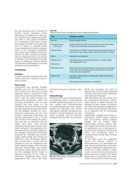
Fig. 4.33 MRI showing an enlarged uterus with<br />
multiple leiomyomas.<br />
Cells diffusely present in most fields examined vs. scattered widely<br />
spaced aggregates of cells<br />
Pleomorphic type: Nuclear pleomorphism appreciated at scanning power<br />
Uniform type: Cells lack pleomorphism but exhibit uniform but marked<br />
nuclear chromatin abnormalities<br />
Expressed in mitotic figures per 10 high power fields in the mitotically<br />
most active areas<br />
Only unequivocal mitotic figures are counted {211}<br />
Nuclei are elongated with blunt or<br />
tapered ends and have finely dispersed<br />
chromatin and small nucleoli. Mitotic figures<br />
usually are infrequent.<br />
Most leiomyomas are more cellular than<br />
the surrounding myometrium. Leiomyomas<br />
lacking increased cellularity are<br />
identified by their nodular circumscription<br />
and by the disorderly arrangement of<br />
the smooth muscle fascicles within them,<br />
out of alignment with the surrounding<br />
myometrium.<br />
Degenerative changes are common in<br />
leiomyomas. Hyaline fibrosis, oedema<br />
and, on occasion, marked hydro p i c<br />
change can be present {525}.<br />
Haemorrhage, necrosis, oedema, myxoid<br />
change, hypercellular foci and cellular<br />
hypertrophy occur in leiomyomas in<br />
women who are pregnant or taking progestins.<br />
Not infre q u e n t l y, there is<br />
increased mitotic activity near the areas<br />
of necrosis.<br />
On the other hand, the coagulative<br />
tumour cell necrosis common in<br />
leiomyosarcoma is not associated very<br />
often with acute inflammation and haemorrhage.<br />
Progestational agents are associated<br />
with a slight increase in mitotic<br />
activity, but not to the level observed in a<br />
leiomyosarcoma. In addition, the mitotic<br />
figures seen in conjunction with inflammatory<br />
necrosis have a normal histologi-<br />
Mesenchymal tumours and related lesions 239


