

Invasive breast carcinoma - IARC
Invasive breast carcinoma - IARC
Invasive breast carcinoma - IARC

You also want an ePaper? Increase the reach of your titles
YUMPU automatically turns print PDFs into web optimized ePapers that Google loves.
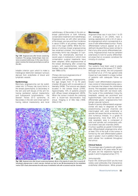
Fig. 1.141 Angiosarcoma after <strong>breast</strong> conserving<br />
treatment. The spongy, haemorrhagic mass contains<br />
occasional solid areas. In this case, the neoplasm<br />
involves the <strong>breast</strong> parenchyma as well as<br />
the skin.<br />
reliable criterion upon which to make a<br />
histological distinction between tumours<br />
derived from endothelia of blood and<br />
lymphatic vessels.<br />
Epidemiology<br />
Mammary angiosarcoma can be subdivided<br />
into 1) Primary (de novo) forms in<br />
the <strong>breast</strong> parenchyma; 2) Secondary in<br />
the skin and soft tissues of the arm following<br />
ipsilateral radical mastectomy<br />
and subsequent lymphoedema - the<br />
S t e w a rt Treves (S-T) syndrome; 3)<br />
Secondary in the skin and chest wall following<br />
radical mastectomy and local<br />
radiotherapy; 4) Secondary in the skin or<br />
b reast parenchyma or both following<br />
conservation treatment and radiotherapy.<br />
Angiosarcomas, as with other sarcomas<br />
of the <strong>breast</strong>, are rare and their incidence<br />
is about 0.05% of all primary malignancies<br />
of the organ {2876}. While the incidence<br />
of primary <strong>breast</strong> angiosarcomas<br />
has remained constant, the incidence of<br />
secondary forms has changed. S-T synd<br />
rome has dramatically declined in<br />
recent years in institutions in which more<br />
conservation surgical treatments have<br />
been adopted, while angiosarcomas of<br />
the <strong>breast</strong> developing after conserving<br />
s u r g e ry with supplementary radiation<br />
therapy have been diagnosed since the<br />
late 1980s {570}.<br />
Primary (de novo) angiosarcoma of<br />
<strong>breast</strong> parenchyma<br />
In patients with primary angiosarcoma,<br />
the age ranges from 17 to 70 years<br />
(median 38 years) with no prevalence of<br />
laterality {2436}. The tumours are deeply<br />
located in the <strong>breast</strong> tissue {2784}.<br />
Approximately 12% of patients present<br />
with diffuse <strong>breast</strong> enlargement {2876}.<br />
When the tumour involves the overlying<br />
skin a bluish-red discoloration may<br />
ensue. Imaging is of little help {1656,<br />
2564,3118}.<br />
Macroscopy<br />
Angiosarcomas vary in size from 1 to 20<br />
cm, averaging 5 cm {2425}, have a<br />
spongy appearance and a rim of vascular<br />
engorgement which corresponds to a<br />
zone of well differentiated tumour. Poorly<br />
differentiated tumours appear as an ill<br />
defined indurated fibrous lesion similar to<br />
that of any other poorly differentiated sarcoma.<br />
Angiosarcomas must be sampled<br />
extensively to look for poorly differentiated<br />
areas that on occasion constitute the<br />
minority of a tumour.<br />
Histopathology<br />
Two systems have been used to grade<br />
angiosarcomas of the <strong>breast</strong> {717,1847}.<br />
Although very similar, the one proposed<br />
by Donnell et al. {717} has gained wide<br />
impact as it was tested in a large number<br />
of patients with adequate follow up<br />
{2436}.<br />
Grade I (well differentiated) angiosarcomas<br />
consist of interanastomosing vascular<br />
channels that dissect the interlobular<br />
stroma. The neoplastic vessels have very<br />
wide lumina filled with red blood cells.<br />
The nuclei of the endothelium lining the<br />
neoplastic vessels are prominent and<br />
hyperchromatic. Care must be taken to<br />
differentiate grade I angiosarcoma from<br />
benign vascular tumours.<br />
Grade III (poorly differentiated) angiosarcomas<br />
are easy to diagnose as interanastomosing<br />
vascular channels are<br />
i n t e rmingled with solid endothelial or<br />
spindle cell areas that show necrotic foci<br />
and numerous mitoses. In a grade III<br />
angiosarcoma, more than 50% of the<br />
total neoplastic area is composed of<br />
solid and spindle cell components without<br />
evident vascular channels {2425}.<br />
A tumour qualifies as grade II (intermediately<br />
differentiated) angiosarcoma when<br />
at least 75% of the bulk of the tumour is<br />
formed by the well differentiated pattern<br />
seen in grade I, but in addition there are<br />
solid cellular foci scattered throughout<br />
the tumour.<br />
Clinical feature<br />
The average age of patients with grade I<br />
angiosarcomas is 43 years while 34 and<br />
29 years are the respective figures for<br />
grade II and III angiosarcomas {717}.<br />
A<br />
B<br />
Fig. 1.142 Angiosarcoma after <strong>breast</strong> conserving treatment. A As in the majority of cases, this is a poorly differentiated<br />
angiosarcoma in which vascular channels are hard to see. B Immunostaining for the endothelial<br />
marker CD31 clearly demonstrates the solid areas of endothelial cells with occasional vascular channels.<br />
Immunoprofile<br />
Factor VIII, CD34 and CD31 are the most<br />
widely used antibodies that characterize<br />
endothelial differentiation. While present<br />
Mesenchymal tumours<br />
95


