

Invasive breast carcinoma - IARC
Invasive breast carcinoma - IARC
Invasive breast carcinoma - IARC

You also want an ePaper? Increase the reach of your titles
YUMPU automatically turns print PDFs into web optimized ePapers that Google loves.
A<br />
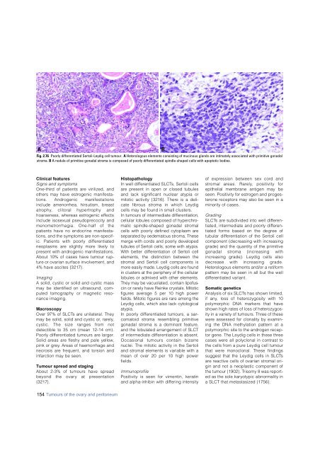
Fig. 2.76 Poorly differentiated Sertoli-Leydig cell tumour. A Heterologous elements consisting of mucinous glands are intimately associated with primitive gonadal<br />
stroma. B A nodule of primitive gonadal stroma is composed of poorly differentiated spindle-shaped cells with apoptotic bodies.<br />
B<br />
Clinical features<br />
Signs and symptoms<br />
O n e - t h i rd of patients are virilized, and<br />
others may have estrogenic manifestations.<br />
Androgenic manifestations<br />
include amenorrhea, hirsutism, bre a s t<br />
a t ro p h y, clitoral hypert rophy and<br />
hoarseness, whereas estrogenic eff e c t s<br />
include isosexual pseudoprecocity and<br />
m e n o m e t rorrhagia. One-half of the<br />
patients have no endocrine manifestations,<br />
and the symptoms are non-specific.<br />
Patients with poorly diff e re n t i a t e d<br />
neoplasms are slightly more likely to<br />
p resent with androgenic manifestations.<br />
About 10% of cases have tumour rupt<br />
u re or ovarian surface involvement, and<br />
4% have ascites {3217}.<br />
I m a g i n g<br />
A solid, cystic or solid and cystic mass<br />
may be identified on ultrasound, computed<br />
tomography or magnetic re s o-<br />
nance imaging.<br />
M a c r o s c o p y<br />
Over 97% of SLCTs are unilateral. They<br />
may be solid, solid and cystic or, rare l y,<br />
cystic. The size ranges from not<br />
detectible to 35 cm (mean 12-14 cm).<br />
Poorly diff e rentiated tumours are larger.<br />
Solid areas are fleshy and pale yellow,<br />
pink or gre y. Areas of haemorrhage and<br />
n e c rosis are frequent, and torsion and<br />
i n f a rction may be seen.<br />
Tumour spread and staging<br />
About 2-3% of tumours have spre a d<br />
beyond the ovary at pre s e n t a t i o n<br />
{ 3 2 1 7 } .<br />
H i s t o p a t h o l o g y<br />
In well diff e rentiated SLCTs, Sertoli cells<br />
a re present in open or closed tubules<br />
and lack significant nuclear atypia or<br />
mitotic activity {3216}. There is a delicate<br />
fibrous stroma in which Leydig<br />
cells may be found in small clusters.<br />
In tumours of intermediate differentiation,<br />
cellular lobules composed of hyperchromatic<br />
spindle-shaped gonadal stromal<br />
cells with poorly defined cytoplasm are<br />
separated by oedematous stroma. These<br />
merge with cords and poorly developed<br />
tubules of Sertoli cells, some with atypia.<br />
With better differentiation of Sertoli cell<br />
elements, the distinction between the<br />
stromal and Sertoli cell components is<br />
more easily made. Leydig cells are found<br />
in clusters at the periphery of the cellular<br />
lobules or admixed with other elements.<br />
They may be vacuolated, contain lipofuscin<br />
or rarely have Reinke crystals. Mitotic<br />
figures average 5 per 10 high power<br />
fields. Mitotic figures are rare among the<br />
Leydig cells, which also lack cytological<br />
atypia.<br />
In poorly diff e rentiated tumours, a sarcomatoid<br />
stroma resembling primitive<br />
gonadal stroma is a dominant feature ,<br />
and the lobulated arrangement of SLCT<br />
of intermediate diff e rentiation is absent.<br />
Occasional tumours contain bizarre<br />
nuclei. The mitotic activity in the Sert o l i<br />
and stromal elements is variable with a<br />
mean of over 20 per 10 high power<br />
f i e l d s .<br />
I m m u n o p ro f i l e<br />
Positivity is seen for vimentin, keratin<br />
and alpha-inhibin with differing intensity<br />
of expression between sex cord and<br />
s t romal areas. Rare l y, positivity for<br />
epithelial membrane antigen may be<br />
seen. Positivity for estrogen and pro g e s-<br />
t e rone receptors may also be seen in a<br />
minority of cases.<br />
G r a d i n g<br />
S L C Ts are subdivided into well diff e re n-<br />
tiated, intermediate and poorly diff e re n-<br />
tiated forms based on the degree of<br />
tubular diff e rentiation of the Sertoli cell<br />
component (decreasing with incre a s i n g<br />
grade) and the quantity of the primitive<br />
gonadal stroma (increasing with<br />
i n c reasing grade). Leydig cells also<br />
d e c rease with increasing grade.<br />
H e t e rologous elements and/or a re t i f o rm<br />
p a t t e rn may be seen in all but the well<br />
d i ff e rentiated variant.<br />
Somatic genetics<br />
Analysis of six SLCTs has shown limited,<br />
if any, loss of heterozygosity with 10<br />
polymorphic DNA markers that have<br />
shown high rates of loss of hetero z y g o s-<br />
ity in a variety of tumours. Three of these<br />
w e re assessed for clonality by examining<br />
the DNA methylation pattern at a<br />
polymorphic site to the androgen re c e p-<br />
tor gene. The Leydig cells in these thre e<br />
cases were all polyclonal in contrast to<br />
the cells from a pure Leydig cell tumour<br />
that were monoclonal. These findings<br />
suggest that the Leydig cells in SLCTs<br />
a re reactive cells of ovarian stromal origin<br />
and not a neoplastic component of<br />
the tumour {1902}. Trisomy 8 was re p o rted<br />
as the sole karyotypic abnormality in<br />
a SLCT that metastasized {1756}.<br />
154 Tumours of the ovary and peritoneum


