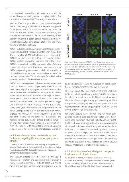
protein-protein interactions. We found several sites forserine/threonine and tyrosine phosphorylation; featuresthat predestine MACC1 as a signal transducer.We identified the gene Met as transcriptional target ofMACC1. Following application the hepatocyte growthfactor (HGF), MACC1 translocates from the cytoplasminto the nucleus, binds to the Met promoter, andinduces its transcription. The HGF/Met pathway is particularlyinvolved in colon cancer metastasis. Thus, weidentified MACC1 as a major regulator of this importantcellular metastasis pathway.MACC1 induces migration, invasion, proliferation, colonyformation, and HGF-mediated scattering in cell culture.These functional MACC1 effects were reverted byMACC1- or Met-specific siRNA. Cells with mutantMACC1 protein interaction domains did neither showMACC1-induced cell motility nor proliferation. Subcutaneous,orthotopic or intrasplenic transplantation ofMACC1-expressing colon cancer cells in mice resulted inelevated tumor growth and increased numbers of distantmetastases. MACC1- or Met-specific shRNA led tolowered numbers of metastases in vivo.MACC1 was overexpressed in human colon cancer comparedto normal tissues. Importantly, MACC1 expressionswere significantly higher in those tumors, thatmetachronously metastasized compared to those,which did not metastasize within up to 10 years. MACC1levels predict the probability of metastasis linked tometastasis-free survival. The correct positive or negativeprediction for metastasis was 74% and 80%, respectively.The 5-year-survival was 80% for patients with lowMACC1, compared to 15% for patients with high MACC1expressions. MACC1 in tumor specimens is an independentprognostic indicator for metastasis andmetastasis-free survival. For clinical practice, MACC1 isan important prognostic gene for early identification ofhigh-risk subjects with colorectal cancer, and a promisingnew target for intervention of metastasis formation.Inhibition of colon cancer metastasis by smallmolecules targeting the metastasis progressorS100A4U. Stein, U. Sack, W. Walther, P.M. Schlag. In cooperationwith W. Birchmeier, I. Fichtner (<strong>MDC</strong>), N. Scudiero, M. Selby(SAIC, Frederick, MD), Robert H. Shoemaker (NationalCancer Institute-Frederick, MD)The calcium-binding protein S100A4 is involved inmetastasis formation and aggressive tumor growth inmany types of cancer. We identified S100A4 as a Wnt/βcatenintarget gene which induces migration, invasionHGF-induced translocation of MACC1 from the cytoplasm into thenucleus in human colon cancer cells, detected by immunofluorescence(bar 50 µm). Stein U, Walther W, Arlt F, Schwabe H, Smith J, Fichtner I,Birchmeier W, Schlag PM. MACC1, a newly identified key regulator ofHGF/Met signaling, predicts colon cancer metastasis. Nature Med15:59-67, 2009and angiogenesis. Hence, its suppression bears potentialfor therapeutic intervention of metastasis.Here we report the identification of small moleculeinhibitors which significantly reduce S100A4 expressionin colorectal carcinoma cells. These inhibitors wereidentified from a high throughput screening of 1,280compounds, employing the S100A4 gene promoterreporter system, and by targeting key molecules of theWnt/β-catenin signaling pathway.Functional assays with selected and validated compoundsrevealed that proliferation rates were diminishedupon treatment while cell viability was only slightlyaffected. More strikingly, migration and invasion ratesof treated cells were significantly decreased by theseinhibitors, but could be rescued by overexpressingS100A4 cDNA. The impact of these small molecules onmetastasis formation in vivo was demonstrated withsmaller and fewer metastases per animal. In summary,our findings present a new strategy to restrict S100A4induced metastasis formation in colon cancer.Clinical application of nonviral gene therapy forlocal treatment of solid tumorsW. Walther, D. Kobelt, R. Siegel, J. Aumann, S. Burock,U. Stein, P. M. Schlag. In cooperation with M. Dietel (Instituteof Pathology, Charité), M. Schleef (PlasmidFactory,Bielefeld), A. Menne (EMS Medical, Nyon, Switzerland)98 Cancer <strong>Research</strong>
For the delivery of naked DNA into cells or tissues agreat variety of procedures is employed in vitro and invivo. Among other physical delivery systems jet-injectionhas developed to an applicable gene transfer technology,which allows gene transfer of small amounts ofnaked DNA into different tissue types with deeper penetrationand improved dispersion.At the Clinic for Surgical Oncology, Charité, Berlin aphase I clinical trial (DeReGe 62) was conducted to evaluatesafety, feasibility and efficiency of intratumoral jetinjectiongene transfer of β-galactosidase (LacZ)-reporter expressing plasmid-DNA into skin metastasesfrom breast cancer and melanoma. Seventeen patientswere enrolled and treated with jet-injection into a singlecutaneous lesion. In the study the safety and efficiencyof the jet-injection gene transfer was shown. Thestudy revealed efficient LacZ mRNA- and proteinexpressionin all treated lesions and rapid clearance ofplasmid-DNA from patient’s blood. The treatment waswell tolerated by all patients and no side effects wereexperienced, indicating clinical applicability of this nonviralapproach for local tumor gene therapy.Use of the minimalistic MIDGE vector forimproved safety, gene transfer and expression inclinical application of gene therapyW. Walther, S. Burock, D. Kobelt, J. Aumann, U. Stein,P. M. Schlag. In cooperation with B. Wittig and M. Schmidt(MOLOGEN AG, Berlin), U. Trefzer (Charité) and I. FichtnerIn result of our clinical gene transfer trial we currentlymake great efforts for further optimization of the nonviralvector by removing unnecessary sequences for theimprovement of transfer- and expression efficiency. Theminimalistic immunologically defined gene expression(MIDGE, Mologen AG, Berlin) vector system provides anonviral linear DNA molecule that is depleted of anybacterial origin, antibiotic resistance sequences or replicationbackbone sequences. This makes MIDGE of highvalue with respect to regulatory requirements for productsafety in clinical applications. Thus, MIDGE vectorsare substantially reduced in size, resulting in highertransfer efficiencies and improved transgene expression.Our in vitro analyses in different human tumor celllines revealed pronounced increase in transfer efficiencyand in transgene expression of the MIDGE vector systemcompared to plasmid-based vectors. The use of thissystem for expression of therapeutic genes, such ashuman TNF-α, mediated high-level expression in vitroand more importantly in vivo and resulted in effectiveantitumoral effects. These expression characteristicsmake MIDGE a good candidate for use in clinical genetherapy applications.Based on our pre-clinical and clinical experiences onnonviral gene transfer a new phase I trial will be initiatedto evaluate safety and efficiency of nonviral jet-injectionapplication of the TNF-α expressing MIDGE-basedvector in patients with skin metastasis from mela -noma. This dose-escalation study will reveal, whetherthe MIDGE-vector is efficiently expressing TNF-α afterthe intratumoral application by jet-injection. It will beimportant to correlate the applied vector dose and theamount of TNF-α expressed in the tumor lesion. Thesafety of the expressed TNF-α and the vector-DNA doseapplied will be examined and will be tested in a clinicalphase II trial, to evaluate the safety and efficacy ofthis approach for the combined modality treatment ofpatients with malignant melanoma.Selected PublicationsStein, U., Walther, W., Arlt, F., Schwabe, H., Smith, J., Fichtner, I., Birchmeier,W., Schlag, P.M. (2009) MACC1, a newly identified key regulator of HGF-MET signaling, predicts colon cancer metastasis. Nat Med. 15, 59-67Fritzmann J, Morkel M, Besser D, Budczies J, Kosel F, Brembeck FH, Stein U,Fichtner I, Schlag PM, Birchmeier W. (2009) A colorectal cancer expressionprofile that includes transforming growth factor beta inhibitor BAMBIpredicts metastatic potential. Gastroenterology 137; 165-175Kemmner W, Wan K, Rüttinger S, Ebert B, Macdonald R, Klamm U, MoestaKT (2008) Silencing of human ferrochelatase causes abundant protoporphyrin-IXaccumulation in colon cancer – a new tool for molecular imaging.FASEB J 22, 500-509Walther W, Siegel R, Kobelt D, Knösel T, Dietel M, Bembenek A, Aumann J,Schleef M, Baier R, Stein U, Schlag PM. (2008) Novel jet-injection technologyfor nonviral intratumoral gene transfer in patients with melanomaand breast cancer. Clin Cancer Res 14, 7545-53.Stein, U., Arlt, F., Walther, W, Smith, J., Waldman, T., Harris, E.D., Mertins,S.D., Heizmann, C.W., Allard, D., Birchmeier, W., Schlag, P.M., and Shoemaker,R.H (2006). The metastasis-associated gene S100A4 is a novel target ofβ-catenin/T-cell factor signalling in colon cancer. Gastroenterology 131,1486-1500.PatentsKemmner W, Schlag PMMicroarray für das Expression Profiling der kompletten zellulärenGlykosylierungsmaschineriePatent application: 9.10.2008Stein U, Lange C, Walther W, Schlag PM.Use of the human LRP/MVP promoter for a vector that can be induced bytherapy.Patent EP 1332219 10.5.2006US 7,297,535 20.11.2007Stein U, Schwabe H, Walther W, Schlag PM.Verwendung des neu-identifizierten Gens 7a5/Prognostin fürTumordiagnostik und Tumortherapie.7a5/Prognostin and its use for tumor diagnostics and therapy.Patent application US 10/564,823 2006Stein U, Walther W, Arlt F, Schlag PM.Methods for diagnosing metastasis by analysing mutations in β-catenin.Patent application US 60/847,547 2006Patent application EP 008333 2007Cancer <strong>Research</strong> 99
- Page 1:
Research Report 2010MAX DELBRÜCK C
- Page 4 and 5:
ContentInhaltContentInhalt.........
- Page 6 and 7:
Surgical OncologyPeter M. Schlag...
- Page 11 and 12:
at the MDC. The role of the institu
- Page 13 and 14:
in discovering genes that contribut
- Page 16 and 17:
The ECRC offers research space and
- Page 18 and 19:
etween disciplines such as biology,
- Page 20 and 21:
approaches from bioinformatics/syst
- Page 23 and 24:
von Humboldt Foundation (AvH). The
- Page 25:
organization to a larger, multi-fac
- Page 28 and 29:
Cardiovascular and Metabolic Diseas
- Page 30 and 31:
electrical signals. More recent wor
- Page 32 and 33:
Basic Cardiovascular FunctionStruct
- Page 34 and 35:
Figure 2: SORLA and sortilin in neu
- Page 36 and 37:
Annette Hammes(Delbrück Fellow)Str
- Page 38 and 39:
Ingo L. MoranoStructure of the Grou
- Page 40 and 41:
Figure 3. Membrane resealing assay
- Page 42 and 43:
Michael GotthardtStructure of the G
- Page 44 and 45:
Structure of the GroupSalim Seyfrie
- Page 46 and 47:
Structure of the GroupFerdinand le
- Page 48 and 49:
Francesca M. SpagnoliStructure of t
- Page 50 and 51:
Structure of the GroupKai M. Schmid
- Page 52 and 53:
Genetics and Pathophysiology of Car
- Page 54 and 55:
Figure 2. Planariato experimentally
- Page 56 and 57:
Norbert HübnerStructure of the Gro
- Page 58 and 59:
Structure of the GroupGroup LeaderF
- Page 60 and 61:
Figure 2. Omega-3 fatty acids prote
- Page 62 and 63:
Structure of the GroupDominik N. M
- Page 64 and 65:
Rainer DietzStructure of the GroupG
- Page 66 and 67:
Figure 2. Cardiac-restricted ablati
- Page 68 and 69:
Ludwig ThierfelderStructure of the
- Page 70 and 71:
standing of the molecular and cellu
- Page 72 and 73:
Structure of the GroupThoralf Niend
- Page 74 and 75: Michael BaderStructure of the Group
- Page 76 and 77: Natriuretic peptide systemJens Butt
- Page 78 and 79: Structure of the GroupZsuzsanna Izs
- Page 80 and 81: Young-Ae LeeStructure of the GroupG
- Page 82 and 83: Structure of the GroupMatthias Selb
- Page 84 and 85: Matthew PoyStructure of the GroupGr
- Page 86 and 87: Jana WolfStructure of the GroupGrou
- Page 88 and 89: Structure of the GroupGroup LeaderD
- Page 91 and 92: Cancer Research ProgramKrebsforschu
- Page 93 and 94: are responsible for the emergence o
- Page 95 and 96: tral component of the canonical Wnt
- Page 97 and 98: lead to an aberrant constitutive ac
- Page 99 and 100: How Notch- and TGFβ signaling casc
- Page 101 and 102: tures of the chronic phase in human
- Page 103 and 104: oped a new safeguard that is based
- Page 105 and 106: Graduate StudentsSeda Cöl ArslanCa
- Page 107 and 108: investigation, as is the cause of c
- Page 109 and 110: Graduate StudentsÖzlem Akilli Özt
- Page 111 and 112: onment, the (cancer) stem cell nich
- Page 113 and 114: The pluripotent state of murine and
- Page 115 and 116: In the morula of the early mouse em
- Page 117 and 118: Graduate StudentsRami Hamscho*Qingb
- Page 119 and 120: esis and granulopoiesis. We showed
- Page 121 and 122: URE ∆/∆ mice regularly develope
- Page 123: PD Dr. Wolfgang Walther (GroupLeade
- Page 127 and 128: ACBDMyc and FoxO transcription fact
- Page 129 and 130: Graduate StudentsKatrin BagolaHolge
- Page 131 and 132: Heinemann, we could also identify t
- Page 133 and 134: Above: Tip of chromosom3L showing t
- Page 135 and 136: Graduate StudentsSarbani Bhattachar
- Page 137 and 138: vesicle transport to the Golgi. Our
- Page 139 and 140: acbFigure 1a: EHD2 is tubulatingpho
- Page 141 and 142: KnowledgeProbabilitiesknownPossible
- Page 143 and 144: Graduate and undergraduatestudentsU
- Page 145 and 146: successive oncogenic mutations. We
- Page 147 and 148: CXCR5 drives the development of ect
- Page 149 and 150: Graduate and undergraduatestudentsW
- Page 151 and 152: Graduate andUndergraduate StudentsM
- Page 153 and 154: Angela MensenStefanie WittstockBjö
- Page 155 and 156: deficient mice, both major effector
- Page 157 and 158: ant of CD3 delta coding for a 45-me
- Page 159 and 160: Robert KudernatschLi-Min LiuAna Mil
- Page 161 and 162: Sebastian GüntherTechnical Assista
- Page 163 and 164: Graduate StudentsJana RolffAnnika W
- Page 165: with murine hepatocytes showed morp
- Page 168 and 169: Function and Dysfunction of the Ner
- Page 170 and 171: The Neuroscience Department also es
- Page 172 and 173: the coming years, Björn Schröder
- Page 174 and 175:
mice. Further analysis of the funct
- Page 176 and 177:
Signaling Pathways and Mechanisms i
- Page 178 and 179:
Control Olig3 -/-ABFigure 2. Geneti
- Page 180 and 181:
Thomas J. JentschStructure of the G
- Page 182 and 183:
Figure 2. Cellular model for ionic
- Page 184 and 185:
Structure of the GroupGroup LeaderF
- Page 186 and 187:
paired-pulse facilitation, less dep
- Page 188 and 189:
Gary R. LewinStructure of the Group
- Page 190 and 191:
Model summarizing the three waves o
- Page 192 and 193:
Structure of the GroupInes Ibañez-
- Page 194 and 195:
Jochen C. MeierStructure of the Gro
- Page 196 and 197:
Björn Christian SchroederStructure
- Page 198 and 199:
Structure of the GroupJan Siemens(S
- Page 200 and 201:
Structure of the GroupGroup LeaderD
- Page 202 and 203:
Imaging of the Living BrainStructur
- Page 204 and 205:
Pathophysiological Mechanisms of Ne
- Page 206 and 207:
Figure 2. Iba1 positive microglia c
- Page 208 and 209:
Erich E. WankerStructure of the Gro
- Page 210 and 211:
Scientific-Technical StaffAnja Frit
- Page 212 and 213:
Structure of the GroupJan Bieschke(
- Page 215 and 216:
Berlin Institute of Medical Systems
- Page 217 and 218:
etes, metabolic diseases and neurod
- Page 219 and 220:
A number of MDC investigators have
- Page 221 and 222:
Technical AssistantsClaudia Langnic
- Page 223 and 224:
has become a standardized data flow
- Page 225:
Phylogeny of cellulase genes from P
- Page 228 and 229:
Experimental and Clinical Research
- Page 230 and 231:
his patients, and a basic research
- Page 232 and 233:
The ultrahigh field MR facility was
- Page 234 and 235:
Structure of the GroupSimone Spuler
- Page 236 and 237:
Ralph KettritzStructure of the Grou
- Page 238 and 239:
Structure of the GroupJeanette Schu
- Page 240 and 241:
Maik GollaschStructure of the Group
- Page 243 and 244:
Technology PlatformsComputational B
- Page 245 and 246:
projects/ard/] to detect repeats li
- Page 247 and 248:
Simulation of line-scan images of C
- Page 249 and 250:
Development of an MRM method for qu
- Page 251 and 252:
mobility or turnover of the underly
- Page 253 and 254:
Left: Inside view of a FACSAria2 (f
- Page 255 and 256:
Examples fort the use of EM methods
- Page 257:
Oviducts lined up in pre-implantati
- Page 260 and 261:
Academic Appointments 2008-2009Beru
- Page 262 and 263:
Buch which is part of the Excellenc
- Page 264 and 265:
“Bioinformatics in Quantitative B
- Page 266 and 267:
Delbrück FellowsDelbrück-Stipendi
- Page 268 and 269:
Yinth Andrea Bernal-Sierra, a PhD s
- Page 270 and 271:
Congresses and Scientific MeetingsK
- Page 272 and 273:
SeminarsSeminare2008Speaker Institu
- Page 274 and 275:
Speaker Institute TitleKiyoshi Mori
- Page 276 and 277:
2009Speaker Institute TitleDavid G.
- Page 278 and 279:
Speaker Institute TitleJuri Rappsil
- Page 280 and 281:
The Helmholtz AssociationDie Helmho
- Page 282 and 283:
The Berlin-Buch CampusDer Campus Be
- Page 284:
the MDC, the existing collaboration
- Page 287 and 288:
Prof. Dr. Gary R. LewinMDC Berlin-B
- Page 289 and 290:
Prof. Dr. Renato ParoCenter of Bios
- Page 291 and 292:
Staff CouncilThe Staff Council is i
- Page 293 and 294:
Type of Financing/Art der Finanzier
- Page 295 and 296:
Research Projects 2008-2009Forschun
- Page 297 and 298:
CIC-5 Regulation und Endocytose am
- Page 299 and 300:
MDCMAX-DELBRÜCK-CENTRUMFÜR MOLEKU
- Page 301 and 302:
Index 275Bröske, A. . . . . . . .
- Page 303 and 304:
Index 277Gross, V. . . . . . . . .
- Page 305 and 306:
Index 279Kur, E. . . . . . . . . .
- Page 307 and 308:
Index 281Piano, F. . . . . . . . .
- Page 309 and 310:
Index 283Smink, J. . . . . . . . .
- Page 312 and 313:
Campus MapCampusplanRobert-Rössle-
- Page 314:
How to find your way to the MDCDer