Research Report 2010 - MDC
Research Report 2010 - MDC
Research Report 2010 - MDC

You also want an ePaper? Increase the reach of your titles
YUMPU automatically turns print PDFs into web optimized ePapers that Google loves.
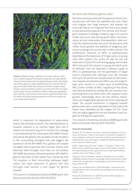
Figure 3. Oligodendrocytic coupling in the corpus callosum of WTmice. Confocal image of the biocytin/streptavidin-Cy3 (red) labellednetwork immunostained for the oligodendroglial markers NG2 (blue)and CNPase (green). A single oligodendrocyte was injected with thegap junction permeable tracer biocytin by whole-cell patch clamp.Biocytin staining revealed a network consisting of 39 cells. The majorityof coupled cells was identified as CNPase expressing oligodendrocytes,only 5% as NG2 positive while 13% was negative for both markers.Within the CNPase positive population 42% of the coupled cellswere weakly expressing NG2.which is important for degradation of extracellularmatrix and promotes invasion. This metalloprotease is,however, released in an inactive, larger form and itneeds to be cleaved to acquire its activity. This cleavageis accomplished by the ectoenzyme MT1-MMP. A factorreleased by the glioma cells activates toll-like receptorsin the surrounding microglial cells and triggers theexpression of the MT1-MMP. Thus, glioma cells exploitmicroglial cells to promote their invasion. Glioma cellsobviously need microglia since they can not produceMT1-MMP themselves: A forced expression MT1-MMP inglioma cells leads to their death. Thus interfering withTLR receptors or their intracellular pathways mightreduce the rapid expansion of glioma cells andmicroglia have become a new target for gliomaresearch.This research is funded by a binational BMBF grant withBozena Kaminska, Warsaw (DLR 01GZ0701).Do stem cells influence glioma cells?We have previously observed that gliomas attract neuralprecursor cells from the subventricular zone. Thesecells migrate over large distances and enwrap thetumor yet they do not originate from the tumor properas was previously suspected. This intrinsic anti-tumorigenicresponse is strongly related to age in an animalmodel and occurs only during youth when neural precursorcells are more active. Consequently, in older animalsthis interaction does not occur. The precursor cellsinhibit tumor growth and addition of exogenous precursorsprolongs the survival rate in older animals. Theproliferative response of NPCs to glioblastomasdepended on the expression of D-type cyclins. In youngmice, NPCs express the cyclins D1 and D2, but theexpression of cyclin D1 is lost during aging, and in adultNPCs only cyclin D2 remains. In young and adult cyclinD2-deficient mice we observed a reduced supply ofNPCs to glioblastomas and the generation of largertumors compared with wild-type mice. We concludethat cyclin D1 and D2 are nonredundant for the antitumorresponse of subventricular NPCs. Loss of a single D-type cyclin results in a smaller pool of proliferatingNPCs, lower number of NPCs migrating to the tumor,and reduced antitumor activity. We are currently characterizingfactors by which stem cells suppress tumorgrowth. Interestingly, there are two distinct mechanisms,one targeting the glioma cell mass inducing celldeath. The second mechanism is targeted towardsglioma stem cells, a small population of cells within thetumor mass identified by the antigen CD 133. Thesecells are highly aggressive and are therefore novel targetsfor therapeutic approaches.This research is funded by a binational BMBF grant withBozena Kaminska, Warsaw (DLR 01GZ0701).Selected publicationsMarkovic, DS., Vinnakota, K, Chirasani, S, Synowitz, M, Raguet, H, Stock, K,Sliwa, M,, Lehmann, S, Kälin, R, van Rooijen, N, Holmbeck, K, Heppner, FL,Kiwit, J, Matyash,V, Lehnardt, S, Kaminska, B, Glass, R, and Kettenmann H.(2009) Glioma induce and exploit microglial MT1-MMP expression fortumor expansion, PNAS, in pressMüller, J, Reyes-Haro, D, Pivneva, T, Nolte, C, Schaette, R, Lübke, J, andKettenmann H. (2009) The principal neurons of the medial nucleus ofthe trapezoid body and NG2+ glial cells receive coordinated excitatorysynaptic input. J Gen. Physiol., in pressCheung, G, Kann, O, Kohsaka, S, Faerber, K, and Kettenmann H. (2009)GABAergic activities enhance macrophage inflammatory protein 1 alpharelease from microglia (brain macrophages) in postnatal mouse brain. JPhysiol. 587,753-768.Hanisch, UK and Kettenmann, H. (2007) Microglia – active sensor andversatile effector cells in the normal and pathologic brain, Nat. Neurosci.10,1387 – 1394.Pocock, JM and Kettenmann H. (2007) Neurotransmitter receptors onmicroglia, Trends Neurosci. 30, 527-535.Function and Dysfunction of the Nervous System 181