Research Report 2010 - MDC
Research Report 2010 - MDC
Research Report 2010 - MDC

Create successful ePaper yourself
Turn your PDF publications into a flip-book with our unique Google optimized e-Paper software.
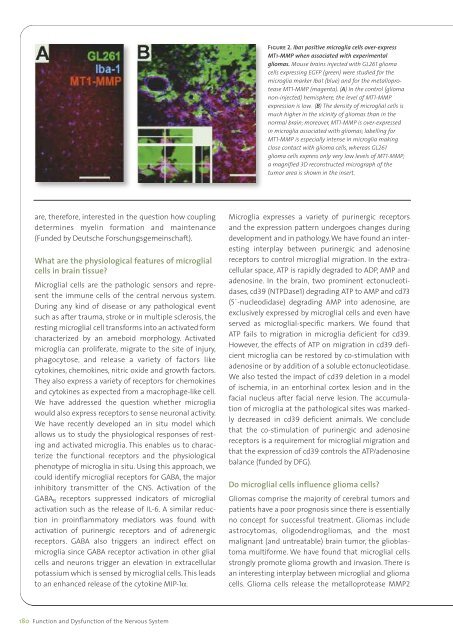
Figure 2. Iba1 positive microglia cells over-expressMT1-MMP when associated with experimentalgliomas. Mouse brains injected with GL261 gliomacells expressing EGFP (green) were studied for themicroglia marker Iba1 (blue) and for the metalloproteaseMT1-MMP (magenta). (A) In the control (gliomanon-injected) hemisphere, the level of MT1-MMPexpression is low. (B) The density of microglial cells ismuch higher in the vicinity of gliomas than in thenormal brain; moreover, MT1-MMP is over-expressedin microglia associated with gliomas; labelling forMT1-MMP is especially intense in microglia makingclose contact with glioma cells, whereas GL261glioma cells express only very low levels of MT1-MMP;a magnified 3D reconstructed micrograph of thetumor area is shown in the insert.are, therefore, interested in the question how couplingdetermines myelin formation and maintenance(Funded by Deutsche Forschungsgemeinschaft).What are the physiological features of microglialcells in brain tissue?Microglial cells are the pathologic sensors and representthe immune cells of the central nervous system.During any kind of disease or any pathological eventsuch as after trauma, stroke or in multiple sclerosis, theresting microglial cell transforms into an activated formcharacterized by an ameboid morphology. Activatedmicroglia can proliferate, migrate to the site of injury,phagocytose, and release a variety of factors likecytokines, chemokines, nitric oxide and growth factors.They also express a variety of receptors for chemokinesand cytokines as expected from a macrophage-like cell.We have addressed the question whether microgliawould also express receptors to sense neuronal activity.We have recently developed an in situ model whichallows us to study the physiological responses of restingand activated microglia. This enables us to characterizethe functional receptors and the physiologicalphenotype of microglia in situ. Using this approach, wecould identify microglial receptors for GABA, the majorinhibitory transmitter of the CNS. Activation of theGABA B receptors suppressed indicators of microglialactivation such as the release of IL-6. A similar reductionin proinflammatory mediators was found withactivation of purinergic receptors and of adrenergicreceptors. GABA also triggers an indirect effect onmicroglia since GABA receptor activation in other glialcells and neurons trigger an elevation in extracellularpotassium which is sensed by microglial cells.This leadsto an enhanced release of the cytokine MIP-1α.Microglia expresses a variety of purinergic receptorsand the expression pattern undergoes changes duringdevelopment and in pathology. We have found an interestinginterplay between purinergic and adenosinereceptors to control microglial migration. In the extracellularspace, ATP is rapidly degraded to ADP, AMP andadenosine. In the brain, two prominent ectonucleotidases,cd39 (NTPDase1) degrading ATP to AMP and cd73(5`-nucleodidase) degrading AMP into adenosine, areexclusively expressed by microglial cells and even haveserved as microglial-specific markers. We found thatATP fails to migration in microglia deficient for cd39.However, the effects of ATP on migration in cd39 deficientmicroglia can be restored by co-stimulation withadenosine or by addition of a soluble ectonucleotidase.We also tested the impact of cd39 deletion in a modelof ischemia, in an entorhinal cortex lesion and in thefacial nucleus after facial nerve lesion. The accumulationof microglia at the pathological sites was markedlydecreased in cd39 deficient animals. We concludethat the co-stimulation of purinergic and adenosinereceptors is a requirement for microglial migration andthat the expression of cd39 controls the ATP/adenosinebalance (funded by DFG).Do microglial cells influence glioma cells?Gliomas comprise the majority of cerebral tumors andpatients have a poor prognosis since there is essentiallyno concept for successful treatment. Gliomas includeastrocytomas, oligodendrogliomas, and the mostmalignant (and untreatable) brain tumor, the glioblastomamultiforme. We have found that microglial cellsstrongly promote glioma growth and invasion. There isan interesting interplay between microglial and gliomacells. Glioma cells release the metalloprotease MMP2180 Function and Dysfunction of the Nervous System