- Page 1:
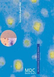
Research Report 2010MAX DELBRÜCK C
- Page 4 and 5:
ContentInhaltContentInhalt.........
- Page 6 and 7:
Surgical OncologyPeter M. Schlag...
- Page 11 and 12:
at the MDC. The role of the institu
- Page 13 and 14:
in discovering genes that contribut
- Page 16 and 17:
The ECRC offers research space and
- Page 18 and 19:
etween disciplines such as biology,
- Page 20 and 21:
approaches from bioinformatics/syst
- Page 23 and 24:
von Humboldt Foundation (AvH). The
- Page 25:
organization to a larger, multi-fac
- Page 28 and 29:
Cardiovascular and Metabolic Diseas
- Page 30 and 31:
electrical signals. More recent wor
- Page 32 and 33:
Basic Cardiovascular FunctionStruct
- Page 34 and 35:
Figure 2: SORLA and sortilin in neu
- Page 36 and 37:
Annette Hammes(Delbrück Fellow)Str
- Page 38 and 39:
Ingo L. MoranoStructure of the Grou
- Page 40 and 41:
Figure 3. Membrane resealing assay
- Page 42 and 43:
Michael GotthardtStructure of the G
- Page 44 and 45:
Structure of the GroupSalim Seyfrie
- Page 46 and 47:
Structure of the GroupFerdinand le
- Page 48 and 49:
Francesca M. SpagnoliStructure of t
- Page 50 and 51:
Structure of the GroupKai M. Schmid
- Page 52 and 53:
Genetics and Pathophysiology of Car
- Page 54 and 55:
Figure 2. Planariato experimentally
- Page 56 and 57:
Norbert HübnerStructure of the Gro
- Page 58 and 59:
Structure of the GroupGroup LeaderF
- Page 60 and 61:
Figure 2. Omega-3 fatty acids prote
- Page 62 and 63:
Structure of the GroupDominik N. M
- Page 64 and 65:
Rainer DietzStructure of the GroupG
- Page 66 and 67:
Figure 2. Cardiac-restricted ablati
- Page 68 and 69:
Ludwig ThierfelderStructure of the
- Page 70 and 71:
standing of the molecular and cellu
- Page 72 and 73:
Structure of the GroupThoralf Niend
- Page 74 and 75:
Michael BaderStructure of the Group
- Page 76 and 77:
Natriuretic peptide systemJens Butt
- Page 78 and 79:
Structure of the GroupZsuzsanna Izs
- Page 80 and 81:
Young-Ae LeeStructure of the GroupG
- Page 82 and 83:
Structure of the GroupMatthias Selb
- Page 84 and 85:
Matthew PoyStructure of the GroupGr
- Page 86 and 87:
Jana WolfStructure of the GroupGrou
- Page 88 and 89:
Structure of the GroupGroup LeaderD
- Page 91 and 92:
Cancer Research ProgramKrebsforschu
- Page 93 and 94:
are responsible for the emergence o
- Page 95 and 96:
tral component of the canonical Wnt
- Page 97 and 98:
lead to an aberrant constitutive ac
- Page 99 and 100:
How Notch- and TGFβ signaling casc
- Page 101 and 102:
tures of the chronic phase in human
- Page 103 and 104:
oped a new safeguard that is based
- Page 105 and 106:
Graduate StudentsSeda Cöl ArslanCa
- Page 107 and 108:
investigation, as is the cause of c
- Page 109 and 110:
Graduate StudentsÖzlem Akilli Özt
- Page 111 and 112:
onment, the (cancer) stem cell nich
- Page 113 and 114:
The pluripotent state of murine and
- Page 115 and 116:
In the morula of the early mouse em
- Page 117 and 118:
Graduate StudentsRami Hamscho*Qingb
- Page 119 and 120:
esis and granulopoiesis. We showed
- Page 121 and 122:
URE ∆/∆ mice regularly develope
- Page 123 and 124:
PD Dr. Wolfgang Walther (GroupLeade
- Page 125 and 126:
For the delivery of naked DNA into
- Page 127 and 128:
ACBDMyc and FoxO transcription fact
- Page 129 and 130:
Graduate StudentsKatrin BagolaHolge
- Page 131 and 132:
Heinemann, we could also identify t
- Page 133 and 134:
Above: Tip of chromosom3L showing t
- Page 135 and 136:
Graduate StudentsSarbani Bhattachar
- Page 137 and 138:
vesicle transport to the Golgi. Our
- Page 139 and 140:
acbFigure 1a: EHD2 is tubulatingpho
- Page 141 and 142:
KnowledgeProbabilitiesknownPossible
- Page 143 and 144:
Graduate and undergraduatestudentsU
- Page 145 and 146:
successive oncogenic mutations. We
- Page 147 and 148:
CXCR5 drives the development of ect
- Page 149 and 150:
Graduate and undergraduatestudentsW
- Page 151 and 152:
Graduate andUndergraduate StudentsM
- Page 153 and 154:
Angela MensenStefanie WittstockBjö
- Page 155 and 156:
deficient mice, both major effector
- Page 157 and 158:
ant of CD3 delta coding for a 45-me
- Page 159 and 160:
Robert KudernatschLi-Min LiuAna Mil
- Page 161 and 162:
Sebastian GüntherTechnical Assista
- Page 163 and 164:
Graduate StudentsJana RolffAnnika W
- Page 165:
with murine hepatocytes showed morp
- Page 168 and 169:
Function and Dysfunction of the Ner
- Page 170 and 171:
The Neuroscience Department also es
- Page 172 and 173:
the coming years, Björn Schröder
- Page 174 and 175:
mice. Further analysis of the funct
- Page 176 and 177:
Signaling Pathways and Mechanisms i
- Page 178 and 179:
Control Olig3 -/-ABFigure 2. Geneti
- Page 180 and 181:
Thomas J. JentschStructure of the G
- Page 182 and 183:
Figure 2. Cellular model for ionic
- Page 184 and 185:
Structure of the GroupGroup LeaderF
- Page 186 and 187:
paired-pulse facilitation, less dep
- Page 188 and 189:
Gary R. LewinStructure of the Group
- Page 190 and 191:
Model summarizing the three waves o
- Page 192 and 193:
Structure of the GroupInes Ibañez-
- Page 194 and 195:
Jochen C. MeierStructure of the Gro
- Page 196 and 197: Björn Christian SchroederStructure
- Page 198 and 199: Structure of the GroupJan Siemens(S
- Page 200 and 201: Structure of the GroupGroup LeaderD
- Page 202 and 203: Imaging of the Living BrainStructur
- Page 204 and 205: Pathophysiological Mechanisms of Ne
- Page 206 and 207: Figure 2. Iba1 positive microglia c
- Page 208 and 209: Erich E. WankerStructure of the Gro
- Page 210 and 211: Scientific-Technical StaffAnja Frit
- Page 212 and 213: Structure of the GroupJan Bieschke(
- Page 215 and 216: Berlin Institute of Medical Systems
- Page 217 and 218: etes, metabolic diseases and neurod
- Page 219 and 220: A number of MDC investigators have
- Page 221 and 222: Technical AssistantsClaudia Langnic
- Page 223 and 224: has become a standardized data flow
- Page 225: Phylogeny of cellulase genes from P
- Page 228 and 229: Experimental and Clinical Research
- Page 230 and 231: his patients, and a basic research
- Page 232 and 233: The ultrahigh field MR facility was
- Page 234 and 235: Structure of the GroupSimone Spuler
- Page 236 and 237: Ralph KettritzStructure of the Grou
- Page 238 and 239: Structure of the GroupJeanette Schu
- Page 240 and 241: Maik GollaschStructure of the Group
- Page 243 and 244: Technology PlatformsComputational B
- Page 245: projects/ard/] to detect repeats li
- Page 249 and 250: Development of an MRM method for qu
- Page 251 and 252: mobility or turnover of the underly
- Page 253 and 254: Left: Inside view of a FACSAria2 (f
- Page 255 and 256: Examples fort the use of EM methods
- Page 257: Oviducts lined up in pre-implantati
- Page 260 and 261: Academic Appointments 2008-2009Beru
- Page 262 and 263: Buch which is part of the Excellenc
- Page 264 and 265: “Bioinformatics in Quantitative B
- Page 266 and 267: Delbrück FellowsDelbrück-Stipendi
- Page 268 and 269: Yinth Andrea Bernal-Sierra, a PhD s
- Page 270 and 271: Congresses and Scientific MeetingsK
- Page 272 and 273: SeminarsSeminare2008Speaker Institu
- Page 274 and 275: Speaker Institute TitleKiyoshi Mori
- Page 276 and 277: 2009Speaker Institute TitleDavid G.
- Page 278 and 279: Speaker Institute TitleJuri Rappsil
- Page 280 and 281: The Helmholtz AssociationDie Helmho
- Page 282 and 283: The Berlin-Buch CampusDer Campus Be
- Page 284: the MDC, the existing collaboration
- Page 287 and 288: Prof. Dr. Gary R. LewinMDC Berlin-B
- Page 289 and 290: Prof. Dr. Renato ParoCenter of Bios
- Page 291 and 292: Staff CouncilThe Staff Council is i
- Page 293 and 294: Type of Financing/Art der Finanzier
- Page 295 and 296: Research Projects 2008-2009Forschun
- Page 297 and 298:
CIC-5 Regulation und Endocytose am
- Page 299 and 300:
MDCMAX-DELBRÜCK-CENTRUMFÜR MOLEKU
- Page 301 and 302:
Index 275Bröske, A. . . . . . . .
- Page 303 and 304:
Index 277Gross, V. . . . . . . . .
- Page 305 and 306:
Index 279Kur, E. . . . . . . . . .
- Page 307 and 308:
Index 281Piano, F. . . . . . . . .
- Page 309 and 310:
Index 283Smink, J. . . . . . . . .
- Page 312 and 313:
Campus MapCampusplanRobert-Rössle-
- Page 314:
How to find your way to the MDCDer


