Diseases and Management of Crops under Protected Cultivation
Diseases and Management of Crops under Protected Cultivation
Diseases and Management of Crops under Protected Cultivation

You also want an ePaper? Increase the reach of your titles
YUMPU automatically turns print PDFs into web optimized ePapers that Google loves.
(<strong>Diseases</strong> <strong>and</strong> <strong>Management</strong> <strong>of</strong> <strong>Crops</strong> <strong>under</strong> <strong>Protected</strong> <strong>Cultivation</strong>)<br />
Use <strong>of</strong> Electron Microscope for Detection <strong>and</strong> Diagnosis <strong>of</strong> Pathogens in<br />
<strong>Protected</strong> <strong>Cultivation</strong><br />
Introduction<br />
Balwinder Singh<br />
Department <strong>of</strong> Anatomy, G.B.P.U.A.&T., Pantnagar - 263145 (Uttarakh<strong>and</strong>)<br />
One <strong>of</strong> the most important tasks in the education <strong>of</strong> a pathologist is learning to distinguish<br />
normal from abnormal tissues. Typically, training<br />
programs provide an adequate background for the<br />
examination <strong>and</strong> interpretation <strong>of</strong> tissues at the<br />
gross <strong>and</strong> light microscopic (lm) levels, leaving the<br />
student pathologist to his/her own devices to<br />
develop necessary skills at the ultrastructural level.<br />
The purpose <strong>of</strong> this presentation is to facilitate<br />
development <strong>of</strong> these skills in ultrastructural<br />
examination/interpretation <strong>of</strong> tissues, by providing a<br />
starting point, some tools for study, direction, <strong>and</strong><br />
finally, a goal at which to aim. Since it would be<br />
unrealistic to attempt to go into depth in the short<br />
time allotted, the presentation will concentrate on<br />
an approach to interpretation <strong>of</strong> ultrastructural<br />
cases while providing a broad overview <strong>of</strong> some<br />
commonly examined tissues.<br />
A human eye can distinguish two points 0.2mm apart. Man’s quest to see the unseen <strong>and</strong><br />
beyond what can be seen with the naked eye led to the discovery <strong>of</strong> simple magnifying glass that<br />
produces an enlarged image <strong>of</strong> an object. Further improvement led to development <strong>of</strong> light<br />
microscopes that use a combination <strong>of</strong> magnifying glasses/lenses. Dr.Ernst Ruska at the<br />
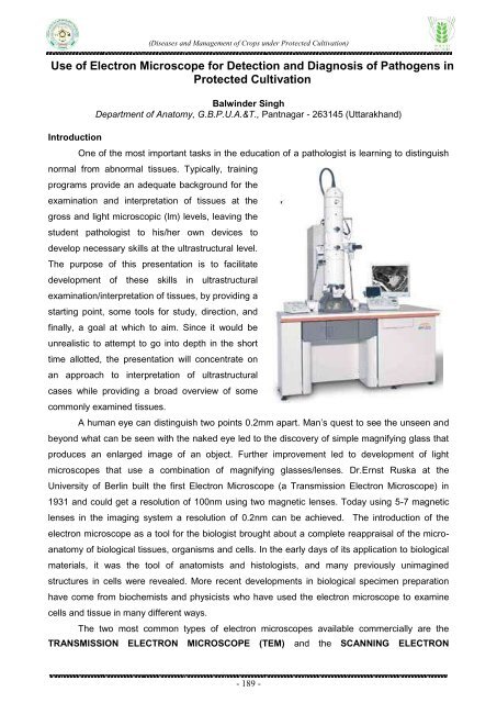
University <strong>of</strong> Berlin built the first Electron Microscope (a Transmission Electron Microscope) in<br />
1931 <strong>and</strong> could get a resolution <strong>of</strong> 100nm using two magnetic lenses. Today using 5-7 magnetic<br />
lenses in the imaging system a resolution <strong>of</strong> 0.2nm can be achieved. The introduction <strong>of</strong> the<br />
electron microscope as a tool for the biologist brought about a complete reappraisal <strong>of</strong> the microanatomy<br />
<strong>of</strong> biological tissues, organisms <strong>and</strong> cells. In the early days <strong>of</strong> its application to biological<br />
materials, it was the tool <strong>of</strong> anatomists <strong>and</strong> histologists, <strong>and</strong> many previously unimagined<br />
structures in cells were revealed. More recent developments in biological specimen preparation<br />
have come from biochemists <strong>and</strong> physicists who have used the electron microscope to examine<br />
cells <strong>and</strong> tissue in many different ways.<br />
The two most common types <strong>of</strong> electron microscopes available commercially are the<br />
TRANSMISSION ELECTRON MICROSCOPE (TEM) <strong>and</strong> the SCANNING ELECTRON<br />
- 189 -