Diseases and Management of Crops under Protected Cultivation
Diseases and Management of Crops under Protected Cultivation
Diseases and Management of Crops under Protected Cultivation

You also want an ePaper? Increase the reach of your titles
YUMPU automatically turns print PDFs into web optimized ePapers that Google loves.
(<strong>Diseases</strong> <strong>and</strong> <strong>Management</strong> <strong>of</strong> <strong>Crops</strong> <strong>under</strong> <strong>Protected</strong> <strong>Cultivation</strong>)<br />
MICROSCOPE (SEM). In the SEM, the specimen is scanned with a focused beam <strong>of</strong> electrons<br />
which produce "secondary" electrons as the beam hits the specimen. These are detected <strong>and</strong><br />
converted into an image on a television screen, <strong>and</strong> a three-dimensional image <strong>of</strong> the surface <strong>of</strong><br />
the specimen is produced. Specimens in the TEM are examined by passing the electron beam<br />
through them, revealing more information <strong>of</strong> the internal structure <strong>of</strong> specimens.<br />
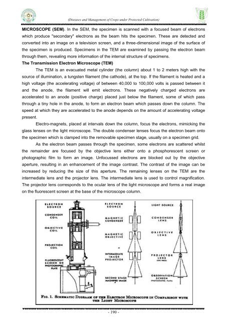
The Transmission Electron Microscope (TEM)<br />
The TEM is an evacuated metal cylinder (the column) about 1 to 2 meters high with the<br />
source <strong>of</strong> illumination, a tungsten filament (the cathode), at the top. If the filament is heated <strong>and</strong> a<br />
high voltage (the accelerating voltage) <strong>of</strong> between 40,000 to 100,000 volts is passed between it<br />
<strong>and</strong> the anode, the filament will emit electrons. These negatively charged electrons are<br />
accelerated to an anode (positive charge) placed just below the filament, some <strong>of</strong> which pass<br />
through a tiny hole in the anode, to form an electron beam which passes down the column. The<br />
speed at which they are accelerated to the anode depends on the amount <strong>of</strong> accelerating voltage<br />
present.<br />
Electro-magnets, placed at intervals down the column, focus the electrons, mimicking the<br />
glass lenses on the light microscope. The double condenser lenses focus the electron beam onto<br />
the specimen which is clamped into the removable specimen stage, usually on a specimen grid.<br />
As the electron beam passes through the specimen, some electrons are scattered whilst<br />
the remainder are focused by the objective lens either onto a phosphorescent screen or<br />
photographic film to form an image. Unfocussed electrons are blocked out by the objective<br />
aperture, resulting in an enhancement <strong>of</strong> the image contrast. The contrast <strong>of</strong> the image can be<br />
increased by reducing the size <strong>of</strong> this aperture. The remaining lenses on the TEM are the<br />
intermediate lens <strong>and</strong> the projector lens. The intermediate lens is used to control magnification.<br />
The projector lens corresponds to the ocular lens <strong>of</strong> the light microscope <strong>and</strong> forms a real image<br />
on the fluorescent screen at the base <strong>of</strong> the microscope column.<br />
- 190 -