- Page 2 and 3:
Pesticide residues in food — 2007
- Page 4:
TABLE OF CONTENTS Page List of part
- Page 7 and 8:
Dr Vicki L. Dellarco, Office of Pes
- Page 10 and 11:
Abbreviations used 3-MC ACTH ADI ai
- Page 12 and 13:
MRL MS MS/MS MTD NMR NOAEC NOAEL NO
- Page 14:
Introduction The toxicological mono
- Page 18 and 19:
AMINOPYRALID First draft prepared b
- Page 20 and 21:
5 higher renal excretion in the gro
- Page 22 and 23:
7 Table 4. Recovery of radioactivit
- Page 24 and 25:
9 Table 7. Pharmacokinetic paramete
- Page 26 and 27:
11 (c) Exposure by inhalation Five
- Page 28 and 29:
13 Table 9. Acute toxicity of amino
- Page 30 and 31:
15 The data from urine analysis rev
- Page 32 and 33:
17 18, monthly for palpable masses.
- Page 34 and 35:
19 Table 12. Body weights of rats f
- Page 36 and 37:
21 Table 15. Results of assays for
- Page 38 and 39:
23 in both groups, feed consumption
- Page 40 and 41:
25 neurotoxicity after 12 months. B
- Page 42 and 43:
27 The same five time-mated NZW rab
- Page 44 and 45:
29 Mortality was increased in all g
- Page 46 and 47:
31 Levels relevant to risk assessme
- Page 48 and 49:
33 References Brooks, K.J. (2001a)
- Page 50 and 51:
35 Consulting, The Dow Chemical Com
- Page 52 and 53:
ATRAZINE First draft prepared by Ru
- Page 54 and 55:
39 in the early 1990s; this reflect
- Page 56 and 57:
41 Maximum concentrations in the bl
- Page 58 and 59:
43 In a study on comparative metabo
- Page 60 and 61:
45 Table 1. Metabolism of atrazine
- Page 62 and 63:
47 Figure 2. Proposed metabolic pat
- Page 64 and 65:
49 to intact skin; and that no read
- Page 66 and 67:
51 the highest dose, and food consu
- Page 68 and 69:
53 mice (evaluated as roughly equiv
- Page 70 and 71:
55 Table 5. Incidence of mammary tu
- Page 72 and 73:
57 Table 6. Selected findings from
- Page 74 and 75:
59 was decreased when compared with
- Page 76 and 77:
61 Table 9. Selected findings of a
- Page 78 and 79:
63 proestrus at the expense of days
- Page 80 and 81:
65 Table 10. Selected studies of ge
- Page 82 and 83:
67 End-point Test object Concentrat
- Page 84 and 85:
69 Table 12. Relevant findings in a
- Page 86 and 87:
71 Table 14. Relevant findings in a
- Page 88 and 89:
73 respectively) and in males at th
- Page 90 and 91:
75 Table 15. Relevant findings in a
- Page 92 and 93:
77 All dams survived until the end
- Page 94 and 95:
79 250 and 500 ppm. Body-weight gai
- Page 96 and 97:
81 In an assay for reverse mutation
- Page 98 and 99:
83 on pubertal development in femal
- Page 100 and 101:
85 parameters (decreased pH, specif
- Page 102 and 103:
87 Table 20. Relevant findings in a
- Page 104 and 105:
89 0, 75, 150 or 300 mg/kg bw per d
- Page 106 and 107:
91 The NOAEL was 25 ppm, equal to 1
- Page 108 and 109:
93 In a study on the effect of atra
- Page 110 and 111:
95 Delayed parturition was seen at
- Page 112 and 113:
97 observed in the pair-fed group.
- Page 114 and 115:
99 examined, offspring in the atraz
- Page 116 and 117:
101 antagonize E2-induced luciferas
- Page 118 and 119:
103 increase in steroidogenesis is
- Page 120 and 121:
105 The immune system of adult mice
- Page 122 and 123:
107 In a later review of cancer epi
- Page 124 and 125:
109 In a 25-day study in rabbits tr
- Page 126 and 127:
111 developmental toxicity were 10
- Page 128 and 129:
113 regulation in male offspring in
- Page 130 and 131:
115 (d) Diaminochlorotriazine (DACT
- Page 132 and 133:
117 Developmental target/critical e
- Page 134 and 135:
119 • The failure to ovulate resu
- Page 136 and 137:
121 The relationship between increa
- Page 138 and 139:
123 Table A2. Comparison of paramet
- Page 140 and 141:
125 Ballantine, L., Murphy, T. G. &
- Page 142 and 143:
127 Dunkelberg, H., Fuchs, J., Heng
- Page 144 and 145:
129 Heneweer, M., van den Berg, M.
- Page 146 and 147:
131 Lindsay, L.A., Wimbert, K.V., G
- Page 148 and 149:
133 Morseth, S.L. (1996d) Chronic (
- Page 150 and 151:
135 Rudzki, M.W., Batastina, G., &
- Page 152 and 153:
137 Tennant, A.H., Peng, B. & Klige
- Page 154 and 155:
AZINPHOS-METHYL First draft prepare
- Page 156 and 157:
141 methylation and oxidation of me
- Page 158 and 159:
143 Table 1. Acute oral toxicity of
- Page 160 and 161:
145 Table 2. Cholinesterase activit
- Page 162 and 163:
147 at the highest dose. In both sp
- Page 164 and 165:
149 Table 6. Cholinesterase activit
- Page 166 and 167:
151 Table 8. Fertility of F 0 and F
- Page 168 and 169:
153 Table 10. Fertility parameters
- Page 170 and 171:
155 Groups of 22 mated Sprague-Dawl
- Page 172 and 173:
157 after completion of the feeding
- Page 174 and 175:
159 reduced motor activity in males
- Page 176 and 177:
161 sensitivity with that of labora
- Page 178 and 179:
163 measures to prevent worker expo
- Page 180 and 181:
165 when brain cholinesterase activ
- Page 182 and 183:
167 Critical end-points for setting
- Page 184 and 185:
169 Eiben, R., Schmidt, W., & Loese
- Page 186 and 187:
171 Myhr, B.C. (1983) Evaluation of
- Page 188 and 189:
LAMBDA-CYHALOTHRIN First draft prep
- Page 190 and 191:
175 Figure 1. Chemical structures o
- Page 192 and 193:
177 In a comparative study, the abs
- Page 194 and 195:
179 Figure 2. Main pathways of biot
- Page 196 and 197:
181 (iv) Dermal sensitization In a
- Page 198 and 199:
183 observed in rats at the highest
- Page 200 and 201:
185 Mortality was not affected by t
- Page 202 and 203:
187 the group at 100 ppm, reduction
- Page 204 and 205:
189 and five females per group were
- Page 206 and 207:
191 10-12%) than those of controls
- Page 208 and 209:
193 Comments Biochemical aspects Or
- Page 210 and 211:
195 Toxicological evaluation Althou
- Page 212 and 213:
197 Metabolism in animals Toxicolog
- Page 214 and 215:
199 Harrison, M.P. (1984a) Cyhaloth
- Page 216 and 217:
DIFENOCONAZOLE First draft prepared
- Page 218 and 219:
203 a sex difference nor any marked
- Page 220 and 221:
205 demonstrated that the highest t
- Page 222 and 223:
207 Figure 3. Proposed metabolic pa
- Page 224 and 225:
209 of the study were unremarkable.
- Page 226 and 227:
211 Table 3. Results of studies of
- Page 228 and 229:
213 The no-observed-adverse-effect
- Page 230 and 231:
215 lower than those of rats in the
- Page 232 and 233:
217 hepatocellular enlargement. In
- Page 234 and 235:
219 i.e. males lost 15.4% and femal
- Page 236 and 237:
221 and 357. This reduced food cons
- Page 238 and 239:
223 There was very high mortality a
- Page 240 and 241:
225 Table 6. Treatment-related hist
- Page 242 and 243:
227 Rats Groups of 80 CRL:CD(SD)®
- Page 244 and 245:
229 Table 7. Histopathology finding
- Page 246 and 247:
231 D. Temporal association. The fe
- Page 248 and 249:
233 2.5 Reproductive toxicity (a) M
- Page 250 and 251:
235 t estes weights in the males at
- Page 252 and 253:
237 continued to be lower than thos
- Page 254 and 255:
239 Corpora lutea in each ovary wer
- Page 256 and 257:
241 S1 + S2, not ossified — —
- Page 258 and 259:
243 in the control group and in the
- Page 260 and 261:
245 physically examined for changes
- Page 262 and 263:
247 Table 16. Results of studies of
- Page 264 and 265:
249 can occur in untreated mice, in
- Page 266 and 267:
251 Study of reproductive toxicity
- Page 268 and 269:
253 Table 19. Results of studies of
- Page 270 and 271:
255 Table 21. Results of studies of
- Page 272 and 273:
257 of the test article on microsom
- Page 274 and 275:
259 Ophthalmoscopic examinations pe
- Page 276 and 277:
261 the radioactivity was re-elimin
- Page 278 and 279:
263 In a single-dose study of neuro
- Page 280 and 281:
265 Estimate of acceptable daily in
- Page 282 and 283:
267 Clapp, M.J.L., Killick, M.E., H
- Page 284 and 285:
269 Herbold, B. (1983c) THS 2212 tr
- Page 286 and 287:
271 Pinto, P. (2006b) Difenoconazol
- Page 288 and 289:
DIMETHOMORPH First draft prepared b
- Page 290 and 291:
275 Five different treatment groups
- Page 292 and 293:
277 To investigate the significance
- Page 294 and 295:
279 and the E : Z isomer ratio was
- Page 296 and 297:
281 Table 10. Tissue distribution o
- Page 298 and 299:
283 (e) Dermal sensitization The de
- Page 300 and 301:
285 females at the highest dose, to
- Page 302 and 303:
287 The NOAEL was 10 mg/kg bw per d
- Page 304 and 305:
289 concentrations in females were
- Page 306 and 307:
291 health, moribundity and mortali
- Page 308 and 309:
293 0-9%, mean, 3.5%; only one out
- Page 310 and 311:
Table 26. Organ weights adjusted to
- Page 312 and 313:
297 cell hyperplasia. The testes tu
- Page 314 and 315:
299 unscheduled DNA synthesis, the
- Page 316 and 317:
301 Figure 3. Cumulative percentage
- Page 318 and 319:
303 Rabbits In a dose-range finding
- Page 320 and 321:
305 (b) Potentiation of hexobarbito
- Page 322 and 323:
307 0.2% or less of the administere
- Page 324 and 325:
309 eye-opening, pinna unfolding or
- Page 326 and 327:
311 Lowest relevant inhalation NOAE
- Page 328 and 329:
313 Gardner, J.R. (1989) CME 151 te
- Page 330:
315 van de Waart, E.J. (1991b) Eval
- Page 333 and 334:
318 Committee on Pesticide residues
- Page 335 and 336:
320 (a) Ocular irritation Rabbits T
- Page 337 and 338:
322 weights were decreased in males
- Page 339 and 340:
324 toxicity was 5 mg/kg bw per day
- Page 341 and 342:
326 respectively; range for histori
- Page 343 and 344: 328 Table 4. Incidence of Leydig-ce
- Page 345 and 346: 330 and/or bilateral) was noted in
- Page 347 and 348: 332 No treatment-related signs of m
- Page 349 and 350: 334 Table 6. Incidence of selected
- Page 351 and 352: 336 or teratogenic potential at the
- Page 353 and 354: 338 and progesterone. The concentra
- Page 355 and 356: 340 the doses used in the 1-year st
- Page 357 and 358: 342 No neurotoxic effects were seen
- Page 359 and 360: 344 Lowest relevant reproductive NO
- Page 361 and 362: 346 Keller, D.A. (1992c) Oncogenici
- Page 364 and 365: PROCYMIDONE First draft prepared by
- Page 366 and 367: 351 Rats Groups of five male and fe
- Page 368 and 369: 353 Table 1. Pharmacokinetic parame
- Page 370 and 371: 355 Seven doses C max (µg equivale
- Page 372 and 373: 357 Three groups of five male and f
- Page 374 and 375: 359 The excretion of radioactivity
- Page 376 and 377: 361 Figure 1. Proposed metabolic pa
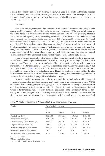
- Page 378 and 379: 363 water consumption were determin
- Page 380 and 381: 365 finding. Histopathology reveale
- Page 382 and 383: 367 The NOAEL was 500 ppm, equivale
- Page 384 and 385: 369 The NOAEL was 100 mg/kg bw per
- Page 386 and 387: 371 at 2000 ppm. Histopathology rev
- Page 388 and 389: 373 b Each test was conducted in tr
- Page 390 and 391: 375 experimental period. Rats were
- Page 392 and 393: 377 hypospadias, testicular atrophy
- Page 396 and 397: 381 The anti-androgenic activities
- Page 398 and 399: 383 but only at 6000 ppm after 13 w
- Page 400 and 401: 385 The results are summarized in T
- Page 402 and 403: 387 males (all litters) in the grou
- Page 404 and 405: 389 procymidone (18-27% of the admi
- Page 406 and 407: 391 Procymidone induced liver tumou
- Page 408 and 409: 393 The Meeting concluded that the
- Page 410 and 411: 395 Toxicologically significant com
- Page 412 and 413: 397 Harada, T. (1983) A review on m
- Page 414 and 415: 399 Murakami, M., Yoshitake, A. & H
- Page 416: 401 Tarui, H. (2005b) In vitro meta
- Page 419 and 420: 404 Explanation Profenofos is the I
- Page 421 and 422: 406 The metabolism of ring-labelled
- Page 423 and 424: 408 2. Toxicological studies 2.1 Ac
- Page 425 and 426: 410 Rabbits Groups of two male and
- Page 427 and 428: 412 Groups of five male and five fe
- Page 429 and 430: 414 control group and in the test g
- Page 431 and 432: 416 of the rabbits at the highest d
- Page 433 and 434: 418 The NOAEL for brain acetylcholi
- Page 435 and 436: 420 Table 2. Histopathological find
- Page 437 and 438: 422 1 out of 70; lowest dose, 3 out
- Page 439 and 440: 424 body‐weight gains (3-10% decr
- Page 441 and 442: 426 treated groups for body-weight
- Page 443 and 444: 428 (b) Short-term studies of neuro
- Page 445 and 446:
430 represented as equally as possi
- Page 447 and 448:
432 pound and the equitoxic mixture
- Page 449 and 450:
434 Inhibition of brain acetylcholi
- Page 451 and 452:
436 Toxicological evaluation Erythr
- Page 453 and 454:
438 Neurotoxicity/delayed neurotoxi
- Page 455 and 456:
440 Harris, S.B. (1982) A teratolog
- Page 457 and 458:
442 Puri, E. (1982a) Autoradiograph
- Page 460 and 461:
PYRIMETHANIL First draft prepared b
- Page 462 and 463:
447 Table 1. Excretion profile in r
- Page 464 and 465:
449 Table 3. Half-life of pyrimetha
- Page 466 and 467:
451 Table 4. Identification of meta
- Page 468 and 469:
453 SN 614 277. The presence of the
- Page 470 and 471:
455 2. Toxicological studies 2.1 Ac
- Page 472 and 473:
457 treated rabbits showed slight e
- Page 474 and 475:
459 still evident in the liver; how
- Page 476 and 477:
461 In a 90-day feeding study, grou
- Page 478 and 479:
463 per day or 800 mg/kg bw per day
- Page 480 and 481:
465 The dosing solutions were prepa
- Page 482 and 483:
467 mortality and morbidity. Change
- Page 484 and 485:
469 2.4 Genotoxicity Pyrimethanil w
- Page 486 and 487:
471 mean body‐weight gains. After
- Page 488 and 489:
473 Analysis of dosing solutions in
- Page 490 and 491:
475 In a 7-day feeding study, group
- Page 492 and 493:
477 at 200 mg/kg bw. These clinical
- Page 494 and 495:
479 s ystemic toxicity was 400 ppm
- Page 496 and 497:
481 Acute toxicity Rat, LD 50 , ora
- Page 498 and 499:
483 Grosshans, F. (2003) Pyrimethan
- Page 500 and 501:
485 Markham, L.P. (1989d) Technical
- Page 502 and 503:
ZOXAMIDE First draft prepared by I.
- Page 504 and 505:
489 The excretion of radioactivity
- Page 506 and 507:
491 Table 3. Mean concentration of
- Page 508 and 509:
493 Table 4. Distribution of metabo
- Page 510 and 511:
495 were also found in the urine, a
- Page 512 and 513:
497 owing to the absence of a dose-
- Page 514 and 515:
499 The NOAEL was 30 000 ppm, equiv
- Page 516 and 517:
501 Table 9. Haematological finding
- Page 518 and 519:
503 Table 11. Body weight, food con
- Page 520 and 521:
505 daily for signs of moribundity,
- Page 522 and 523:
507 Table 14 Results of studies of
- Page 524 and 525:
509 phase. The study was certified
- Page 526 and 527:
511 Table 16. Spleen weights and hi
- Page 528 and 529:
513 FOB/motor activity assessment,
- Page 530 and 531:
515 Excretion was primarily in the
- Page 532 and 533:
517 days 14 to 21. Increased relati
- Page 534 and 535:
519 Lowest relevant inhalation NOAE
- Page 536 and 537:
521 Morrison, R.D. & Gillette, D.M.
- Page 538 and 539:
ANNEX 1 Reports and other documents
- Page 540 and 541:
525 31. Pesticide residues in food:
- Page 542 and 543:
527 67. Pesticide residues in food
- Page 544:
529 101. Pesticide residues in food