Third Day Poster Session, 17 June 2010 - NanoTR-VI
Third Day Poster Session, 17 June 2010 - NanoTR-VI
Third Day Poster Session, 17 June 2010 - NanoTR-VI

You also want an ePaper? Increase the reach of your titles
YUMPU automatically turns print PDFs into web optimized ePapers that Google loves.
P<br />
P<br />
P<br />
P<br />
P<br />
<strong>Poster</strong> <strong>Session</strong>, Thursday, <strong>June</strong> <strong>17</strong><br />
Theme F686 - N1123<br />
Fabrication of Aligned Silk Fibroin Nanofibers by Electrospinning<br />
1<br />
2<br />
3<br />
4<br />
Gamze DoanP P, UGüldemet BaalUP P*, Ali Bora BaltaP P, Ouz BayraktarP<br />
1 Department of Textile Engineering, Uak University, Uak 64100, Turkey<br />
2<br />
PDepartment of Textile Engineering, Ege University, zmir 35100, Turkey<br />
PDepartment of Biotechnology and Bioengineering, zmir Istitute of Technology, zmir 35430, Turkey<br />
4<br />
PDepartment of Chemical Engineering, zmir Istitute of Technology, zmir 35430, Turkey<br />
3<br />
Abstract- Aligned nanofibers provide some advantages in fabrication of scaffolds for tissue engineering. In this study, silk fibroin nanofibers<br />
were fabricated by an electrospinning unit with a rotating drum as a collector at three different drum speeds (surface velocity), and the influence<br />
of drum speed on fiber size and alignment were investigated.<br />
Tissue engineering is a field of regenerative medicine,<br />
which deals with the development of tissue substitutes<br />
(scaffolds) to repair, maintain, or improve the function of<br />
diseased or damaged tissues. Mimicking of cell<br />
microenviroment as close as possible when designing<br />
scaffolds is the key issue in tissue engineering [1].<br />
Electrospun biopolymer nanofibers have potential uses as<br />
scaffolds, due to their resemblance to natural extra cellular<br />
matrix (ECM), high surface area to volume ratio and high<br />
porosities [2]. The ECM is a nano fibrous network which<br />
holds cells and tissues together and provides a controlled<br />
environment inside which migratory cells can move and<br />
interact with each other [3].<br />
Several natural or synthetic biodegrable polymers have been<br />
turned into scaffolds for tissue engineering. Silk fibroin (SF)<br />
is a great candidate for this purpose. It has a slow degradation<br />
rate, good mechanical properties, high oxygen permeability<br />
and it is non-toxic [4, 5].<br />
One of the most widely used method for the fabrication of<br />
nanofibrous scaffolds is electrospinning. This method involves<br />
the ejection and stretching of a polymer solution or melt from<br />
a capillary tube by electrostatic forces. In electrospinning<br />
method, stationary collectors are used for the production of<br />
random nanofiber bundles. Rotating targets such as disks and<br />
drums are used for the fabrication of aligned nanofibers [6, 7].<br />
Alignment of nanofibers plays an important role in repairing<br />
tissues that have structural orientation in one direction such as<br />
muscle and nerve tissues. According to contact guidance<br />
theory aligned nanofiber scaffolds can exhibit more ECM<br />
production than random nanofiber scaffolds [8]. These aligned<br />
nanofiber scaffolds also have a more dense structure and high<br />
strength value compared to random nanofiber scaffolds [9].<br />
In this study, aligned nanofibers were fabricated from silk<br />
fibroin (SF) by utilizing an electrospinning set up with a<br />
rotating drum and the effects of the surface velocity of the<br />
rotating drum on fiber size and alignment of fibers were<br />
investigated.<br />
Silk fibroin solution was prepared using 98% formic acid.<br />
The concentration of SF in the solution was 6 wt%. Applied<br />
voltage was 20 kV. Flow rate was set to 7 μL/min. Distance<br />
between the collector and the needle tip was adjusted to 11.2<br />
cm. All of these parameters were kept constant, except the<br />
surface velocity of the rotating drum. Electrospinning was<br />
performed at three different surface velocities: 50, 100 and<br />
150m/min. Results revealed that the influence of drum speed<br />
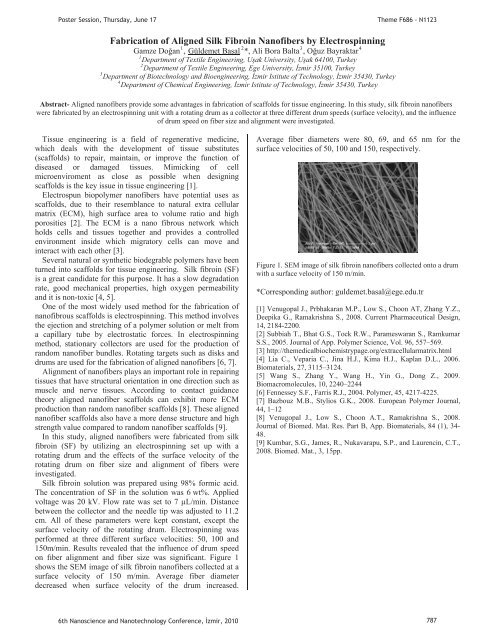
on fber alignment and fber size was significant. Figure 1<br />
shows the SEM image of silk fibroin nanofibers collected at a<br />
surface velocity of 150 m/min. Average fiber diameter<br />
decreased when surface velocity of the drum increased.<br />
Average fiber diameters were 80, 69, and 65 nm for the<br />
surface velocities of 50, 100 and 150, respectively.<br />
Figure 1. SEM image of silk fibroin nanofibers collected onto a drum<br />
with a surface velocity of 150 m/min.<br />
*Corresponding author: guldemet.basal@ege.edu.tr<br />
[1] Venugopal J., Prbhakaran M.P., Low S., Choon AT, Zhang Y.Z.,<br />
Deepika G., Ramakrishna S., 2008. Current Pharmaceutical Design,<br />
14, 2184-2200.<br />
[2] Subbiah T., Bhat G.S., Tock R.W., Parameswaran S., Ramkumar<br />
S.S., 2005. Journal of App. Polymer Science, Vol. 96, 557–569.<br />
[3] http://themedicalbiochemistrypage.org/extracellularmatrix.html<br />
[4] Lia C., Veparia C., Jina H.J., Kima H.J., Kaplan D.L., 2006.<br />
Biomaterials, 27, 3115–3124.<br />
[5] Wang S., Zhang Y., Wang H., Yin G., Dong Z., 2009.<br />
Biomacromolecules, 10, 2240–2244<br />
[6] Fennessey S.F., Farris R.J., 2004. Polymer, 45, 42<strong>17</strong>-4225.<br />
[7] Bazbouz M.B., Stylios G.K., 2008. European Polymer Journal,<br />
44, 1–12<br />
[8] Venugopal J., Low S., Choon A.T., Ramakrishna S., 2008.<br />
Journal of Biomed. Mat. Res. Part B, App. Biomaterials, 84 (1), 34-<br />
48.<br />
[9] Kumbar, S.G., James, R., Nukavarapu, S.P., and Laurencin, C.T.,<br />
2008. Biomed. Mat., 3, 15pp.<br />
6th Nanoscience and Nanotechnology Conference, zmir, <strong>2010</strong> 787