Third Day Poster Session, 17 June 2010 - NanoTR-VI
Third Day Poster Session, 17 June 2010 - NanoTR-VI
Third Day Poster Session, 17 June 2010 - NanoTR-VI

You also want an ePaper? Increase the reach of your titles
YUMPU automatically turns print PDFs into web optimized ePapers that Google loves.
<strong>Poster</strong> <strong>Session</strong>, Thursday, <strong>June</strong> <strong>17</strong><br />
Theme F686 - N1123<br />
Optical and Structural Characterization of Y 2 O 3 :Nd 3+ Phosphors via Thermal Decomposition Method<br />
G. Bilir* and G. Özen<br />
Department of Physics, stanbul Technical University, Maslak-stanbul 34469, Turkey<br />
Abstract: The Y 2 O 3 :Nd 3+ nanophosphors were synthesized by using thermal decomposition method. The powders were annealed at different<br />
temperatures to investigate annealing temperature dependence of the crystallite size. Average particle size of the products were calculated by<br />
using Scherrer Formula from the X-ray diffractograms Luminescence measurement were performed for all samples at room temperature. Also<br />
SEM/EDS measurements were confirmed the calculated particle sizes from XRD diffractograms.<br />
Materials with nanostructure attracted considerable attention<br />
because of potential applications in optoelectronics and<br />
photonics[1-4]. Phosphor materials find wide applications<br />
ranging from fluorescent lamp to luminescence immunoassay.<br />
These materials essentially convert one type of energy into<br />
visible radiation and hence, phosphor materials are called<br />
optical transducer[5].<br />
In this work nanosized Y 2 O 3 samples doped with x=0.2, 0.5,<br />
1, 2, 5, 10 mol% Nd 3+ ions (Y 2-x Nd x O 3 ) were prepared by<br />
thermal decomposition of yttrium-neodymium alginate.<br />
Obtained products were annealed at 600, 800 and 1000 to<br />
investigate particle size dependence on annealing temperature.<br />
X-ray diffraction investigations were carried out with<br />
Philips TM model(Cu-K) diffractometer at 40 kV in the 2 range<br />
from 20 o to 60 o . Also SEM images of the samples were taken by<br />
using JEOL 6335F model scanning electron microscope(SEM).<br />
Both of XRD and SEM measurements show that the particle<br />
sizes of Y 2 O 3 :Nd 3+ samples were ranging from 20nm to 40 nm<br />
which are consistent with the values reported in literature[6].<br />
Representative X-ray diffractograms and SEM images are given<br />
in Figs.1-2.<br />
Princeton Instruments SP2500i model monochromator and<br />
Acton series ID441-C Model InGaAs detector for the<br />
detection.<br />
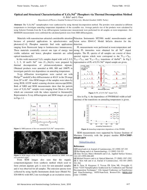
PL measurements were performed at room temperature and<br />
strong PL intensities were obtained for all Nd 3+ doped<br />
samples. The PL spectra of all samples are consist of three<br />
spectral regions which were correspond to the 4 F 3/2 4 I 9/2 ,<br />
4 F 3/2 4 I 11/2 and 4 F 3/2 4 I 13/2 transitions of theNd 3+ . In Fig.2<br />
representative of PL of 0.5% Nd 3+ doped sample are given.<br />
Figure 3: PL of 0.5% Nd 3+ doped Y 2O 3<br />
Also in Fig. 4, the dependence of FWHM(full width at half<br />
maxima) of the transitions on annealing temperature is given.<br />
Figure 1: XRD patterns of the non-annealed Y 2O 3:Nd 3+ nanopowders<br />
Figure 2: SEM images of the %0,5Nd doped Y 2O 3 annealed at 1000C (left) and<br />
non-annealed (right)<br />
From SEM images also seen that the organic<br />
component(alginate) from synthesis method which used to<br />
form yttrium alginate gels is exist for non-annealed samples<br />
and it was observed for all samples. The emission spectra were<br />
collected by using Apollo Instruments diode laser (Model No:<br />
S30-808-6) with 805.2 nm wavelength as an excitation source,<br />
Figure 4:Annealing temperature dependence of the FWHM<br />
SEM measurements were supported by Science Institute of<br />
Marmara University with the project number FEN-CDRP-<br />
090409-0079<br />
*bilirg@itu.edu.tr<br />
References<br />
[1]Promod and et al, Journal of Luminescence, 82 (1999) 187-193<br />
[2] Hai Huang and et al, Nanotechnology, 13 (2002) 318-323<br />
[3] Gino Tessari, Marco Bettinelli and et al., Applied Surface Science,<br />
144-145 (1999) 686-689<br />
[4] Michael Nazarov and et al, Optical Materials, 27 (2005) 1587-1592<br />
[5] T. Kim Anh and et al, Journal of Luminescence, 102-103 (2003)<br />
391-394<br />
[6] D. Tatar, H. Kaygusuz, F. Tezcan, FB. Erim, ML. Oveçolu, G.<br />
Ozen “Y2O3 Nanophosphors Synthesized by Combustion and Thermal<br />
Decomposition Techniques” 11th Annual Conference on NanoScience<br />
and NanoTechnology, NSTI-NanoTech, Boston-USA (MO81-919)<br />
(<strong>June</strong> 1-5/2008)<br />
6th Nanoscience and Nanotechnology Conference, zmir, <strong>2010</strong> 6<strong>17</strong>