

Photonic crystals in biology
Photonic crystals in biology
Photonic crystals in biology

Create successful ePaper yourself
Turn your PDF publications into a flip-book with our unique Google optimized e-Paper software.
Poster Session, Tuesday, June 15<br />
Theme A1 - B702<br />
Absorption Hypothesis: Attachment of Beetles to Nanoporous Substrates<br />
Elena Gorb 1 *, Solveig Kle<strong>in</strong>z 1 and Stanislav Gorb 1<br />
1 Department of Functional Morphology and Biomechanics, Zoological Institute, University of Kiel, 24098 Kiel, Germany<br />
Abstract-Traction experiments with ladybird beetles showed that forces on nanoporous substrates were significantly lower t hat those on solid<br />
surface samples. The comparison of the evolution <strong>in</strong> contact angles of the two fluids, polar water and non-polar m<strong>in</strong>eral oil, showed that porous<br />
substrates absorbed both polar and non-polar fluids, whereas solid surface samples did not. Thus, the reduction of <strong>in</strong>sect attachment on<br />
nanoporous surfaces may be expla<strong>in</strong>ed by (1) the absorption of the secretion fluid from <strong>in</strong>sect adhesive pads by porous media and/or (2) the<br />
effect of surface roughness.<br />
It has been repeatedly reported that micro- and<br />
nanostructured waxy surfaces of plants strongly reduce <strong>in</strong>sect<br />
attachment. To expla<strong>in</strong> anti-adhesive properties of such<br />
substrates, four hypotheses have been previously proposed: (a)<br />
roughness hypothesis; (b) contam<strong>in</strong>ation hypothesis; (c) fluid<br />
absorption hypothesis; and (d) wax dissolv<strong>in</strong>g hypothesis [1].<br />
Recently, only the two first hypotheses ([2 – 6] for (a) and [7 –<br />
9] fo r (b)) were proven. To date, the wax d issolv<strong>in</strong>g<br />
hypothesis and the fluid absorption hypothesis have not been<br />
experimentally tested. In the present study, we used<br />
nanoporous substrates with the same pore diameter (220 – 235<br />
nm), but different porosity (area of voids <strong>in</strong> a material surface,<br />
normalized over the total area), <strong>in</strong> order to test the fluid<br />
absorption hypothesis, claim<strong>in</strong>g that the structured wax<br />
coverage of plants may absorb the fluid from the setal surface<br />
of <strong>in</strong>sect adhesive pads and by this reduce the adhesion force.<br />
We performed traction force measurements with tethered<br />
seven-spotted ladybird beetles Cocc<strong>in</strong>ella septempunctata L.<br />
(Coleoptera, Cocc<strong>in</strong>ellidae), walk<strong>in</strong>g on five different<br />
substrates [10]: (1) smooth glass plate; (2) smooth solid Al 2 O 3<br />
(sapphire) disc; (3 – 5) three types of nanoporous Al 2 O 3 discs<br />
(back side of anodisc membranes Whatman, Schleicher and<br />
Schuell, Whatman International Ltd., Maidstone, UK) hav<strong>in</strong>g<br />
the porosity of 28, 42 and 51%. Forces were measured with a<br />
load cell force transducer (10g capacity, Biopac Systems Ltd.,<br />
Santa Barbara, CA, USA). Both males (n=10) and females<br />
(n=10) were used <strong>in</strong> the experiments.<br />
We found that the forces ranged from 0.16 to 16.59 mN <strong>in</strong><br />
males and from 0.32 to 8.99 mN <strong>in</strong> females. The highest force<br />
values were obta<strong>in</strong>ed on the smooth surfaces, where males<br />
generated considerably higher forces compared to females. On<br />
all three porous substrates, the forces were significantly<br />
reduced, and the only difference was obta<strong>in</strong>ed between<br />
nanoporous membranes hav<strong>in</strong>g the highest and lowest<br />
porosity. Males produced essentially lower forces than<br />
females on porous samples [10].<br />
The reduction of <strong>in</strong>sect attachment on nanoporous<br />
substrates may be expla<strong>in</strong>ed by (1) possible absorption of the<br />
secretory fluid from <strong>in</strong>sect pads by porous media and (2)<br />
surface roughness, reduc<strong>in</strong>g real contact area between tenent<br />
setae of <strong>in</strong>sect adhesive pads and substrate.<br />
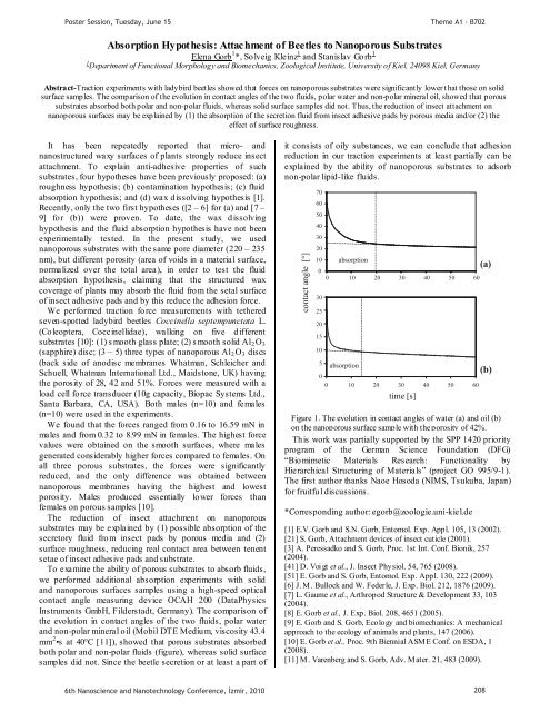
To exam<strong>in</strong>e the ability of porous substrates to absorb fluids,<br />
we performed additional absorption experiments with solid<br />
and nanoporous surfaces samples us<strong>in</strong>g a high-speed optical<br />
contact angle measur<strong>in</strong>g device OCAH 200 (DataPhysics<br />
Instruments GmbH, Filderstadt, Germany). The comparison of<br />
the evolution <strong>in</strong> contact angles of the two fluids, polar water<br />
and non-polar m<strong>in</strong>eral oil (Mobil DTE Medium, viscosity 43.4<br />
mm 2 •s at 40°C [11]), showed that porous substrates absorbed<br />
both polar and non-polar fluids (figure), whereas solid surface<br />
samples did not. S<strong>in</strong>ce the beetle secretion or at least a part of<br />
it consists of oily substances, we can conclude that adhesion<br />
reduction <strong>in</strong> our traction experiments at least partially can be<br />
expla<strong>in</strong>ed by the ability of nanoporous substrates to adsorb<br />
non-polar lipid-like fluids.<br />
contact angle [ ° ]<br />
70<br />
60<br />
50<br />
40<br />
30<br />
20<br />
10<br />
0<br />
0 10 20 30 40 50 60<br />
30<br />
25<br />
20<br />
15<br />
10<br />
5<br />
absorption<br />
absorption<br />
0<br />
0 10 20 30 40 50 60<br />
time [s]<br />
(a)<br />
(b)<br />
Figure 1. The evolution <strong>in</strong> contact angles of water (a) and oil (b)<br />
on the nanoporous surface sample with the porosity of 42%.<br />
This work was partially supported by the SPP 1420 priority<br />
program of the German Science Foundation (DFG)<br />
“Biomimetic Materials Research: Functionality by<br />
Hierarchical Structur<strong>in</strong>g of Materials” (project GO 995/9-1).<br />
The first author thanks Naoe Hosoda (NIMS, Tsukuba, Japan)<br />
for fruitful discussions.<br />
*Correspond<strong>in</strong>g author: egorb@zoologie.uni-kiel.de<br />
[1] E.V. Gorb and S.N. Gorb, Entomol. Exp. Appl. 105, 13 (2002).<br />
[21] S. Gorb, Attachment devices of <strong>in</strong>sect cuticle (2001).<br />
[3] A. Peressadko and S. Gorb, Proc. 1st Int. Conf. Bionik, 257<br />
(2004).<br />
[41] D. Voi gt et al., J. Insect Physiol. 54, 765 (2008).<br />
[51] E. Gorb and S. Gorb, Entomol. Exp. Appl. 130, 222 (2009).<br />
[6] J. M. Bullock and W. Federle, J. Exp. Biol. 212, 1876 (2009).<br />
[7] L. Gaume et al., Arthropod Structure & Development 33, 103<br />
(2004).<br />
[8] E. Gorb et al., J. Exp. Biol. 208, 4651 (2005).<br />
[9] E. Gorb and S. Gorb, Ecology and biomechanics: A mechanical<br />
approach to the ecology of animals and plants, 147 (2006).<br />
[10] E. Gorb et al., Proc. 9th Biennial ASME Conf. on ESDA, 1<br />
(2008).<br />
[11] M. Varenberg and S. Gorb, Adv. Mater. 21, 483 (2009).<br />
6th Nanoscience and Nanotechnology Conference, zmir, 2010 208


