Photonic crystals in biology
Photonic crystals in biology
Photonic crystals in biology
Create successful ePaper yourself
Turn your PDF publications into a flip-book with our unique Google optimized e-Paper software.
Poster Session, Tuesday, June 15<br />
Theme A1 - B702<br />
Electros pun Hybrid Scaffolds for Bone Tissue Repair<br />
1 *<br />
1 urkey.<br />
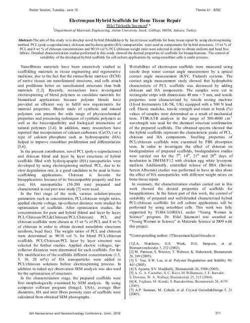
Abstract-The aim of this study is to develop novel hybrid (blend&layer by layer) tissue scaffolds for bone tissue repair by us<strong>in</strong>g electrosp<strong>in</strong>n<strong>in</strong>g<br />
method. PCL (poly--caprolactone), chitosan and hydroxyapatite (HA) nanoparticles were used as components for hybrid structures. 15 wt % of<br />
PCL and 8 wt % of chitosan concentrations and 90/10 vol % PCL/chitosan weight ratio were selected <strong>in</strong> order to obta<strong>in</strong> uniform and bead free<br />
fabrics. Detailed characterization studies performed <strong>in</strong> this study showed the desired properties of scaffolds for bioapplications. The control of<br />
suitability of the developed hybrid scaffolds for cell culture applications by us<strong>in</strong>g osteoblast cells is under process.<br />
Nanofibrous materials have been extensively studied as<br />
scaffold<strong>in</strong>g materials <strong>in</strong> tissue eng<strong>in</strong>eer<strong>in</strong>g and regenerative<br />
medic<strong>in</strong>e, due to the fact that the extracellular matrices (ECM)<br />
of native tissues are nanofeatured structures, and cells attach<br />
and proliferate better on nanofeatured structures than bulk<br />
materials [1,2]. Recently, researchers have <strong>in</strong>vestigated<br />
electrosp<strong>in</strong>n<strong>in</strong>g of blend polymers as candidate materials for<br />
biomedical applications because polymer blends have<br />
provided an efficient way to fulfill new requirements for<br />
material properties. Blends made of synthetic and natural<br />
polymers can present the wide range of physicochemical<br />
properties and process<strong>in</strong>g techniques of synthetic polymers as<br />
well as the biocompatibility and biological <strong>in</strong>teractions of<br />
natural polymers [3,4]. In addition, many researchers have<br />
reported that <strong>in</strong>corporation of calcium carbonate (CaCO 3 ) or a<br />
type of calcium phosphate such as hydroxyapatite (HA)<br />
helped to improve osteoblast proliferation and differentiation<br />
[5,6].<br />
In the present contribution, novel PCL (poly--caprolactone)<br />
and chitosan blend and layer by layer structures of hybrid<br />
scaffolds filled with hydroxyapapite (HA) nanoparticles were<br />
developed by us<strong>in</strong>g electrosp<strong>in</strong>n<strong>in</strong>g method. PCL, due to its<br />
slow degradation rate, is a good candidate to be used <strong>in</strong> bonescaffold<strong>in</strong>g<br />
applications. Chitosan is favorite for<br />
bioapplications because of its biocompatible property and low<br />
cost. HA nanoparticles (50-200 nm) prepared and<br />
characterized <strong>in</strong> our previous study [7] were used.<br />
In the first stage of the work, various solution/process<br />
parameters such as concentration, PCL/chitosan weight ratios,<br />
applied electric voltage, tip-collector distance were studied for<br />
optimization of scaffolds. After optimization studies, the<br />
concentrations for pure and hybrid (blend and layer by layer;<br />
PCL/Chitosan/PCL&Chitosan/PCL/Chitosan) PCL and<br />
chitosan scaffolds were chosen as 15 wt % of PCL and 8 wt %<br />
of chitosan <strong>in</strong> order to obta<strong>in</strong> desired nanofabric structures<br />
(uniform, bead free). The weight ratios of PCL and chitosan<br />
were determ<strong>in</strong>ed as 90/10 vol % for blend PCL/chitosan<br />
scaffolds. PCL/Chitosan/PCL layer by layer structure was<br />
selected for further studies. Applied electric voltages, tipcollector<br />
distances were determ<strong>in</strong>ed for each scaffolds. For the<br />
HA modification of the scaffolds different concentrations (1.5,<br />
5, 10, 20 wt%) of HA nanoparticles were added to<br />
PCL/chitosan solutions before electrosp<strong>in</strong>n<strong>in</strong>g process. In<br />
addition to naked eye observation SEM analysis was also used<br />
for the optimization of structures.<br />
In the characterization stage, the prepared scaffolds were<br />
first morphologically exam<strong>in</strong>ed by SEM analysis. By us<strong>in</strong>g<br />
computer software program (ImageJ, USA), average fiber<br />
diameters, HA and <strong>in</strong>ter fibers porosity sizes of scaffolds were<br />
calculated from obta<strong>in</strong>ed SEM photographs.<br />
Wettabilities of electrospun scaffolds were measured us<strong>in</strong>g<br />
sessile drop water contact angle measurement by a optical<br />
contact angle measurement (KSV, F<strong>in</strong>land) systems. The<br />
contact angle measurement study showed that hydrophobic<br />
characteristic of PCL scaffolds was decreased by add<strong>in</strong>g<br />
chitosan and HA components. The samples were cut <strong>in</strong><br />
rectangular strips with dimensions 40 mm × 5 mm, and tensile<br />
properties were characterized by tensile test<strong>in</strong>g mach<strong>in</strong>e<br />
(Llyod Instruments LK-5K, UK) equipped with a 500 N load<br />
cell. Elastic modulus, tensile strength and stra<strong>in</strong> at break (%)<br />
values of samples were determ<strong>in</strong>ed as a result of mechanical<br />
tests. FTIR-ATR analysis <strong>in</strong> the range of 500-4000 cm -1<br />
wavelength was used for the chemical structure confirmation<br />
of the prepared scaffolds. The obta<strong>in</strong>ed spectra showed that<br />
the hybrid scaffolds represent the characteristic peaks of PCL,<br />
chitosan and HA components. Swell<strong>in</strong>g properties of<br />
PCL/chitosan scaffolds were exam<strong>in</strong>ed by PBS absorption<br />
tests. In order to <strong>in</strong>vestigate the effect of chitosan on<br />
biodegradation of prepared scaffolds, biodegradation studies<br />
were carried out for the 7 th , 14 th , 21 st<br />
and 28 th<br />
days of<br />
<strong>in</strong>cubation <strong>in</strong> DMEM/F12 with chicken egg white lysozyme<br />
medium. Additionally, controlled release of BSA (Bov<strong>in</strong>e<br />
Serum Album<strong>in</strong>) studies was performed to have an idea about<br />
the effect of HA nanoparticles with different weight ratios on<br />
bone tissue repair.<br />
In summary, the characterization studies carried out <strong>in</strong> this<br />
work showed the desired properties of scaffolds for<br />
bioapplications. In the future part of this work, the control of<br />
suitability of prepared and well/detailed characterized hybrid<br />
PCL/chitosan scaffolds for cell culture applications will be<br />
performed by us<strong>in</strong>g osteoblast cells. This work was fully<br />
supported by TUBA/LOREAL under “Young Women <strong>in</strong><br />
Science” program. Dr. <br />
“Young Woman <strong>in</strong> Science” <strong>in</strong> Materials Science at 2009 with<br />
this project.<br />
*Correspond<strong>in</strong>g author: 1Thtsasmazel@atilim.edu.tr<br />
[1]J.A. Matthews, G.E. Wnek, D.G. Simpson, et al.<br />
Biomacromolecules 3, 232 (2002).<br />
[2] M. Pattison, S. Wurster, T. Webster, K. Haberstroh, Biomaterials<br />
26, 249 (2005).<br />
[3] Y. You, S.W. Lee, et al. Polymer Degradation and Stability 90,<br />
441 (2005).<br />
[4] S. Aparna, S.V. Madihally, Biomaterials 26, 5500 (2005).<br />
[5] A. G. A. Coombes, S. C. Rizzi, M.Williamson, J. E. Barralet,<br />
S. Downes, W. A. Wallace, Biomaterials 25, 315 (2004).<br />
[6] K. Fujihara, M. Kotaki, S. Ramakrishna, Biomaterials 26, 4139<br />
(2005).<br />
[7] A.P. Sommer, M. Çehreli, et al. Crystal Growth&Design 5, 21<br />
(2005).<br />
6th Nanoscience and Nanotechnology Conference, zmir, 2010 211


