- Page 2 and 3:
Medical modelling
- Page 4 and 5:
Medical modelling The application o
- Page 6 and 7:
Contents Preface ix Acknowledgement
- Page 8:
Contents vii 6.10 Rehabilitation ap
- Page 11 and 12:
x Preface Therefore, it is hoped th
- Page 14 and 15:
1.1 Background 1 Introduction The p
- Page 16 and 17:
Introduction 3 Whilst this book is
- Page 18 and 19:
Long axis 1.1 The anatomical positi
- Page 20 and 21:
Introduction 7 1.3 The major refere
- Page 22 and 23:
Medical imaging for rapid prototypi
- Page 24 and 25:
Medical imaging for rapid prototypi
- Page 26 and 27:
2.2.3 Anatomical coverage Medical i
- Page 28 and 29:
Medical imaging for rapid prototypi
- Page 30 and 31:
2.6 X-ray scatter artefact in a CT
- Page 32 and 33:
2.9 Close up of noise. Medical imag
- Page 34 and 35:
2.12 Smooth data. Medical imaging f
- Page 36 and 37:
Medical imaging for rapid prototypi
- Page 38 and 39:
Medical imaging for rapid prototypi
- Page 40 and 41:
1 Medical imaging for rapid prototy
- Page 42 and 43:
Medical imaging for rapid prototypi
- Page 44 and 45:
Medical imaging for rapid prototypi
- Page 46 and 47:
Export data format and media 33 (NE
- Page 48 and 49:
3.3 Media Export data format and me
- Page 50 and 51:
4.1 Pixel data operations 4 Working
- Page 52 and 53:
4.3 Effect of a high threshold. 4.4
- Page 54 and 55:
Working with medical scan data 41 4
- Page 56 and 57:
Working with medical scan data 43 4
- Page 58 and 59:
Working with medical scan data 45 S
- Page 60 and 61:
4.4 Two-dimensional formats Working
- Page 62 and 63:
Working with medical scan data 49 4
- Page 64 and 65:
Working with medical scan data 51 I
- Page 66 and 67:
4.18 Close up view showing facets.
- Page 68 and 69:
Working with medical scan data 55 4
- Page 70 and 71:
Working with medical scan data 57 4
- Page 72 and 73:
5.1 Background to rapid prototyping
- Page 74 and 75:
Physical reproduction 61 developing
- Page 76 and 77:
Physical reproduction 63 will displ
- Page 78 and 79:
Physical reproduction 65 As there i
- Page 80 and 81:
Physical reproduction 67 However, t
- Page 82 and 83:
Data quality Physical reproduction
- Page 84 and 85:
Physical reproduction 71 In some ca
- Page 86 and 87:
Physical reproduction 73 Overhangin
- Page 88 and 89:
Physical reproduction 75 5.10 SL mo
- Page 90 and 91:
Physical reproduction 77 5.12 Selec
- Page 92 and 93: 5.13 A Perfactory ® model of a man
- Page 94 and 95: 5.15 FDM TM model of the mandible.
- Page 96 and 97: Table 5.3 Advantages and disadvanta
- Page 98 and 99: Physical reproduction 85 In medical
- Page 100 and 101: Table 5.5 Advantages and disadvanta
- Page 102 and 103: 5.7 Jetting head technology 5.7.1 P
- Page 104 and 105: 5.23 ThermoJet ® model of the mand
- Page 106 and 107: 5.24 LOM TM model of the mandible.
- Page 108 and 109: 5.26 LOM TM model of a partial face
- Page 110 and 111: 6 Case studies The following case s
- Page 112 and 113: IMPLEMENTATION 6.1 Implementation c
- Page 114 and 115: Post model Hospital department Pati
- Page 116 and 117: Case studies 103 When using this so
- Page 118 and 119: Case studies 105 (Mimics). Image ma
- Page 120 and 121: Case studies 107 full co-operation
- Page 122 and 123: Case studies 109 communication and
- Page 124 and 125: • to achieve patient’s agreemen
- Page 126 and 127: 6.2.4 Rapid prototyping technologie
- Page 128 and 129: Case studies 115 not be suitable. A
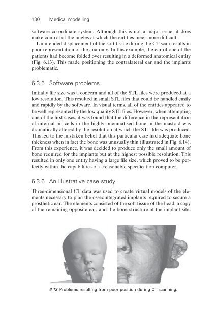
- Page 130 and 131: Case studies 117 scale of the data
- Page 132 and 133: 6.7 SL model with signifi cant stai
- Page 134 and 135: Case studies 121 6.9a Smooth surfac
- Page 136 and 137: Case studies 123 slice editing of t
- Page 138 and 139: Case studies 125 6.12 Removal of bo
- Page 140 and 141: Case studies 127 18. Williams R J,
- Page 144 and 145: Case studies 131 6.14 The effect of
- Page 146 and 147: Case studies 133 6.18 The block ove
- Page 148 and 149: Case studies 135 create template (3
- Page 150 and 151: 6.4.3 Materials and methods Case st
- Page 152 and 153: 6.22 Resected bone compared to SLA
- Page 154 and 155: Case studies 141 9. Robinson R P, C
- Page 156 and 157: Case studies 143 multi-plane reform
- Page 158 and 159: Case studies 145 6.25 The custom ti
- Page 160 and 161: Case studies 147 6.28 Plain radiogr
- Page 162 and 163: Case studies 149 reproduction of ex
- Page 164 and 165: Case studies 151 manner. The softwa
- Page 166 and 167: Case studies 153 surface on which t
- Page 168 and 169: 6.33 The guide being fi tted to the
- Page 170 and 171: Case studies 157 The more fundament
- Page 172 and 173: Case studies 159 25. Sarment D P, A
- Page 174 and 175: 6.37 Stereolithography models. 6.38
- Page 176 and 177: Case studies 163 6.41 Merged pre- a
- Page 178 and 179: REHABILITATION APPLICATIONS 6.8 Reh
- Page 180 and 181: 6.8.3 Methods Preliminary trial of
- Page 182 and 183: Case studies 169 prototyping system
- Page 184 and 185: Case studies 171 The aperture for t
- Page 186 and 187: Case studies 173 7. Manners C R (19
- Page 188 and 189: Case studies 175 during the acquisi
- Page 190 and 191: 6.46 Smoothing the data (exaggerate
- Page 192 and 193:
6.49 Plaster fi lled SL mould. Case
- Page 194 and 195:
Case studies 181 diffi cult for hos
- Page 196 and 197:
Case studies 183 extremely diffi cu
- Page 198 and 199:
Case studies 185 back as shown in F
- Page 200 and 201:
6.54 The result of the Boolean subt
- Page 202 and 203:
6.56 The SLA model of the plate. Ca
- Page 204 and 205:
6.59 SLA implant on SLA model of de
- Page 206 and 207:
Case studies 193 In the near future
- Page 208 and 209:
Case studies 195 anatomical forms a
- Page 210 and 211:
Case studies 197 Establishing the c
- Page 212 and 213:
Case studies 199 6.63 The rough (a)
- Page 214 and 215:
Case studies 201 The fi nal prosthe
- Page 216 and 217:
Case studies 203 grating the techno
- Page 218 and 219:
Case studies 205 6.12 Rehabilitatio
- Page 220 and 221:
Case studies 207 • Data capture N
- Page 222 and 223:
Case studies 209 placement of two i
- Page 224 and 225:
Case studies 211 technique that has
- Page 226 and 227:
Case studies 213 6.72 The bar locat
- Page 228 and 229:
Case studies 215 Stereolithography
- Page 230 and 231:
Case studies 217 authors intend to
- Page 232 and 233:
Case studies 219 17. Kau C H, Zhuro
- Page 234 and 235:
Case studies 221 310, Sainte-Foy, Q
- Page 236 and 237:
6.77a The physically surveyed cast.
- Page 238 and 239:
Creation of relief Case studies 225
- Page 240 and 241:
6.81 Construction curves. Case stud
- Page 242 and 243:
6.83 The support structure in 3D Li
- Page 244 and 245:
Finishing Case studies 231 The cast
- Page 246 and 247:
Case studies 233 6.14 Rehabilitatio
- Page 248 and 249:
6.86 The RPD framework designed in
- Page 250 and 251:
Second experiment Case studies 237
- Page 252 and 253:
Case studies 239 6.90 Close up view
- Page 254 and 255:
Table 6.2 Process steps and associa
- Page 256 and 257:
Case studies 243 9. Budtz-Jorgensen
- Page 258 and 259:
Case studies 245 micro-computed tom
- Page 260 and 261:
Case studies 247 supports became av
- Page 262 and 263:
Case studies 249 However, the probl
- Page 264 and 265:
6.96 Model of one of the samples su
- Page 266 and 267:
6.15.9 Reference Case studies 253 1
- Page 268 and 269:
Case studies 255 mummies. Mimics so
- Page 270 and 271:
Case studies 257 6.101 3D reconstru
- Page 272 and 273:
Case studies 259 6.103 The stages o
- Page 274 and 275:
6.16.5 Conclusions Case studies 261
- Page 276 and 277:
Case studies 263 the patient’s de
- Page 278 and 279:
Case studies 265 and sharp radii re
- Page 280 and 281:
Suitable technologies identifi ed C
- Page 282 and 283:
Case studies 269 6.107 A close-up p
- Page 284 and 285:
6.110 The selected region of facial
- Page 286 and 287:
Case studies 273 patterns were buil
- Page 288 and 289:
Case studies 275 7. Cheah C M, Chua
- Page 290 and 291:
Future developments 277 slices to b
- Page 292 and 293:
Future developments 279 ments that
- Page 294 and 295:
Glossary and explanatory notes 281
- Page 296 and 297:
Glossary and explanatory notes 283
- Page 298 and 299:
Further reading on anatomy Last’s
- Page 300 and 301:
Fundamentals of Facial Prosthetics
- Page 302 and 303:
Rapid Prototyping and Tooling Resea
- Page 304 and 305:
Contacts 3D Systems 26081 Avenue Ha
- Page 306 and 307:
Marcam Engineering GmbH Fahrenheits
- Page 308 and 309:
3D Lightyear TM 229, 247, 249-51, 2
- Page 310:
Removable Partial Denture 219-20, 2
- Page 313 and 314:
4.10 A three-dimensional shaded ima
- Page 315:
6.41 Merged pre- and post-operative