Create successful ePaper yourself
Turn your PDF publications into a flip-book with our unique Google optimized e-Paper software.
POSTER: ACUTE WOUNDS<br />
Poster: Acute Wounds<br />
P 204<br />
A NEW TREATMENT IN THE SKIN LESIONS DUE TO RADIATION THERAPY<br />
AnnaMaria Ippolito 1 , Ornella Forma 2 , Alessandro Corsi 1 , Paolo Cuffaro 1 ,<br />
Roberto Cassino 1<br />
1 Vulnera – Italian Vulnological Center (Turin, Italy);<br />
2 San Raffaele Hospital (Milan, Italy).<br />
Aim: The most common side effects of oncologic radiotherapy are rash or redness,<br />
permanent pigmentation, and scarring in the treated area (radiodermatitis). Radiation<br />
therapy causes inflammation of tissues and organs in and around the body site radiated:<br />
for example, radiation can inflame skin to cause a burn. Aim of the work is to<br />
demonstrate the effectiveness of a new product containing glycerosomas carrying<br />
hyaluronate whose mechanism of action is to decrease local inflammation, to form<br />
protective barrier and to regenerate skin.<br />
Methods: The study involved 20 patients with skin damages due to oncologic radiation<br />
therapy. The treatment protocol provides local treatment consisting of applying a small<br />
amount of the product on the radiodermatitis, after cleansing with neutral wet wipes; no<br />
secondary dressing needed. The renewal of the dressing was provided every 24 hours.<br />
The effectiveness evaluation was based on the removal of clinical signs of inflammation<br />
and the reduction of pain, evaluated with VAS (Visual Analogue Scale).<br />
Results: We had pain reduction in more than 70% of patients and effectiveness in terms<br />
of improvement of the skin condition in 100% of cases. The mean time of treatment was<br />
about 2 months, but the pain reduction has been achieved within 3 weeks.<br />
Conclusions: The treatment of radiodermatitis has always been quite empirical:<br />
clinicians suggest to most of patients a nonspecific treatment with steroidal creams or<br />
burns product. Now we think to have a specific product that can become part of an<br />
effective protocol to prevent and care the skin damages of radiotherapy.<br />
P 205<br />
Poster: Acute Wounds<br />
TREATMENT OF GRADE II AND III RADIODERMATITIS IN CANCER PATIENTS<br />
UNDERGOING RADIOTHERAPY HEAD AND NECK<br />
Roselie Corcini Pinto 1 , Bianca BortoliI Souza 1 , Karina Zanella Arrosi 1 ,<br />
Fabiane Mendonça da Rosa 1 , Elaine Cristina Costa 1 , Leila Maria de Abreu Jaggi 1 ,<br />
Neiro Waechter da Motta 1<br />
1 Serviço de Radioterapia/Hospital Santa Rita da Irmandade Santa Casa de Misericórdia<br />
de Porto Alegre (Porto Alegre, Brazil).<br />
Introduction: Radiotherapy uses radioisotopes for cancer treatment leading the<br />
malignant cells to lose their clonogenicity through the deleterious effects of radioactivity<br />
on the tissues. In this process, the lining epithelial cells, are hit or radiodermatitis<br />
triggering skin lesions that are decisive for the therapeutic outcome. In the literature,<br />
there is no evidence of a protocol on the effectiveness of the products listed for the<br />
recovery of radiodermatitis. Being at the discretion of the caregivers stopping treatment,<br />
analgesics and anti-inflammatory, healing frequency, use of salt solutions, antibiotics and<br />
sprays sulfadiazinas among others.<br />
Objective: To evaluate the efficacy of antimicrobial hydrofiber overburden of comprised<br />
of sodium carboxymethyl cellulose and ionic silver for treating radiodermatitis with<br />
grades II and III, in respect to the time of healing, pain relief and manipulation.<br />
Methods: A prospective cohort pilot prognosis of 20 patients with grades II and III<br />
radiodermatitis using the cover of carboxymethylcellulose and silver. Patients were<br />
monitored, guided and evaluated by nurses as the application of visual scale of pain,<br />
vital signs, number of dressings made and appearance of the lesion.<br />
Results/Conclusions: 20 of the analyzed patients, we had, pain relief in 98% in the first<br />
24 hours and 2% in 48 hours, 100% used analgesics in the first 24 hours and 3% in 48<br />
hours, 85% heal an exchange of dressings, and 15% had 2, 55% healed in 6 days, 35%<br />
(5) 5% (9) and 5% (10). Thus we conclude that there was a positive response to<br />
treatment with significant reduction of pain, reduction of injuries in an average of eight<br />
days, and a reduction in the number of interventions without compromising the safety of<br />
the treatment. They also emphasized the importance of the intervention of the nurse,<br />
education and therapeutic decisions in these patients, opening a precedent for a<br />
randomized clinical trial that is already in underway.<br />
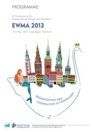
<strong>EWMA</strong> <strong>2013</strong><br />
COPENHAGEN<br />
15-17 May · <strong>2013</strong><br />
Danish Wound<br />
Healing Society<br />
131