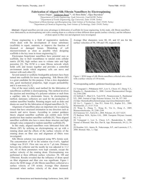
PPPPPPoster Session, Thursday, June 17Theme F686 - N1123Fabrication of Aligned Silk Fibro<strong>in</strong> Nanofibers by Electrosp<strong>in</strong>n<strong>in</strong>g1234Gamze DoanP P, UGüldemet BaalUP P*, Ali Bora BaltaP P, Ouz BayraktarP1 Department of Textile Eng<strong>in</strong>eer<strong>in</strong>g, Uak University, Uak 64100, Turkey2PDepartment of Textile Eng<strong>in</strong>eer<strong>in</strong>g, Ege University, zmir 35100, TurkeyPDepartment of Biotechnology and Bioeng<strong>in</strong>eer<strong>in</strong>g, zmir Istitute of Technology, zmir 35430, Turkey4PDepartment of Chemical Eng<strong>in</strong>eer<strong>in</strong>g, zmir Istitute of Technology, zmir 35430, Turkey3Abstract- Aligned nanofibers provide some advantages <strong>in</strong> fabrication of scaffolds for tissue eng<strong>in</strong>eer<strong>in</strong>g. In this study, silk fibro<strong>in</strong> nanofiberswere fabricated by an electrosp<strong>in</strong>n<strong>in</strong>g unit with a rotat<strong>in</strong>g drum as a collector at three different drum speeds (surface velocity), and the <strong>in</strong>fluenceof drum speed on fiber size and alignment were <strong>in</strong>vestigated.Tissue eng<strong>in</strong>eer<strong>in</strong>g is a field of regenerative medic<strong>in</strong>e,which deals with the development of tissue substitutes(scaffolds) to repair, ma<strong>in</strong>ta<strong>in</strong>, or improve the function ofdiseased or damaged tissues. Mimick<strong>in</strong>g of cellmicroenviroment as close as possible when design<strong>in</strong>gscaffolds is the key issue <strong>in</strong> tissue eng<strong>in</strong>eer<strong>in</strong>g [1].Electrospun biopolymer nanofibers have potential uses asscaffolds, due to their resemblance to natural extra cellularmatrix (ECM), high surface area to volume ratio and highporosities [2]. The ECM is a nano fibrous network whichholds cells and tissues together and provides a controlledenvironment <strong>in</strong>side which migratory cells can move and<strong>in</strong>teract with each other [3].Several natural or synthetic biodegrable polymers have beenturned <strong>in</strong>to scaffolds for tissue eng<strong>in</strong>eer<strong>in</strong>g. Silk fibro<strong>in</strong> (SF)is a great candidate for this purpose. It has a slow degradationrate, good mechanical properties, high oxygen permeabilityand it is non-toxic [4, 5].One of the most widely used method for the fabrication ofnanofibrous scaffolds is electrosp<strong>in</strong>n<strong>in</strong>g. This method <strong>in</strong>volvesthe ejection and stretch<strong>in</strong>g of a polymer solution or melt froma capillary tube by electrostatic forces. In electrosp<strong>in</strong>n<strong>in</strong>gmethod, stationary collectors are used for the production ofrandom nanofiber bundles. Rotat<strong>in</strong>g targets such as disks anddrums are used for the fabrication of aligned nanofibers [6, 7].Alignment of nanofibers plays an important role <strong>in</strong> repair<strong>in</strong>gtissues that have structural orientation <strong>in</strong> one direction such asmuscle and nerve tissues. Accord<strong>in</strong>g to contact guidancetheory aligned nanofiber scaffolds can exhibit more ECMproduction than random nanofiber scaffolds [8]. These alignednanofiber scaffolds also have a more dense structure and highstrength value compared to random nanofiber scaffolds [9].In this study, aligned nanofibers were fabricated from silkfibro<strong>in</strong> (SF) by utiliz<strong>in</strong>g an electrosp<strong>in</strong>n<strong>in</strong>g set up with arotat<strong>in</strong>g drum and the effects of the surface velocity of therotat<strong>in</strong>g drum on fiber size and alignment of fibers were<strong>in</strong>vestigated.Silk fibro<strong>in</strong> solution was prepared us<strong>in</strong>g 98% formic acid.The concentration of SF <strong>in</strong> the solution was 6 wt%. Appliedvoltage was 20 kV. Flow rate was set to 7 μL/m<strong>in</strong>. Distancebetween the collector and the needle tip was adjusted to 11.2cm. All of these parameters were kept constant, except thesurface velocity of the rotat<strong>in</strong>g drum. Electrosp<strong>in</strong>n<strong>in</strong>g wasperformed at three different surface velocities: 50, 100 and150m/m<strong>in</strong>. Results revealed that the <strong>in</strong>fluence of drum speedon fber alignment and fber size was significant. Figure 1shows the SEM image of silk fibro<strong>in</strong> nanofibers collected at asurface velocity of 150 m/m<strong>in</strong>. Average fiber diameterdecreased when surface velocity of the drum <strong>in</strong>creased.Average fiber diameters were 80, 69, and 65 nm for thesurface velocities of 50, 100 and 150, respectively.Figure 1. SEM image of silk fibro<strong>in</strong> nanofibers collected onto a drumwith a surface velocity of 150 m/m<strong>in</strong>.*Correspond<strong>in</strong>g author: guldemet.basal@ege.edu.tr[1] Venugopal J., Prbhakaran M.P., Low S., Choon AT, Zhang Y.Z.,Deepika G., Ramakrishna S., 2008. Current Pharmaceutical Design,14, 2184-2200.[2] Subbiah T., Bhat G.S., Tock R.W., Parameswaran S., RamkumarS.S., 2005. Journal of App. Polymer Science, Vol. 96, 557–569.[3] http://themedicalbiochemistrypage.org/extracellularmatrix.html[4] Lia C., Veparia C., J<strong>in</strong>a H.J., Kima H.J., Kaplan D.L., 2006.Biomaterials, 27, 3115–3124.[5] Wang S., Zhang Y., Wang H., Y<strong>in</strong> G., Dong Z., 2009.Biomacromolecules, 10, 2240–2244[6] Fennessey S.F., Farris R.J., 2004. Polymer, 45, 4217-4225.[7] Bazbouz M.B., Stylios G.K., 2008. European Polymer Journal,44, 1–12[8] Venugopal J., Low S., Choon A.T., Ramakrishna S., 2008.Journal of Biomed. Mat. Res. Part B, App. Biomaterials, 84 (1), 34-48.[9] Kumbar, S.G., James, R., Nukavarapu, S.P., and Laurenc<strong>in</strong>, C.T.,2008. Biomed. Mat., 3, 15pp.6th Nanoscience and Nanotechnology Conference, zmir, 2010 787
PPPPPoster Session, Thursday, June 17Theme F686 - N1123Electrosp<strong>in</strong>nability of Hyaluronic Acid123UGamze DoanUP P*, Güldemet BaalP P, Ali Bora BaltaP P, Ouz BayraktarP1PDepartment of Textile Eng<strong>in</strong>eer<strong>in</strong>g, Uak University, Uak 64100, Turkey2PDepartment of Textile Eng<strong>in</strong>eer<strong>in</strong>g, Ege University, zmir 35100, TurkeyPDepartment of Biotechnology and Bioeng<strong>in</strong>eer<strong>in</strong>g, zmir Istitute of Technology, zmir 35430, Turkey4PDepartment of Chemical Eng<strong>in</strong>eer<strong>in</strong>g, zmir Istitute of Technology, zmir 35430, Turkey3Abstract- Natural biopolymer nanofibers have advantages <strong>in</strong> tissue eng<strong>in</strong>eer<strong>in</strong>g applications due to their good biocompatibility,biodegradability, and resemblance to native extracellular matrix which enhances the tissue regeneration. Hyaluronic acid, a naturalbiopolymer exist<strong>in</strong>g <strong>in</strong> human body, is commonly used <strong>in</strong> scaffold fabrication. One recent fabrication technique for the creation of scaffoldsis electrosp<strong>in</strong>n<strong>in</strong>g. However, electrosp<strong>in</strong>ability of hyaluronic acid is very poor due its high viscosity. This study reveals the problems faced <strong>in</strong>electrosp<strong>in</strong>n<strong>in</strong>g of hyaluronic acid and focuses on determ<strong>in</strong><strong>in</strong>g proper solvent systems and blends which allow the successful production ofhyaluronic acid nanofibers.4Hyaluronic acid attracts much attention <strong>in</strong> tissueeng<strong>in</strong>eer<strong>in</strong>g applications s<strong>in</strong>ce it is a basic component ofextra cellular matrix [1]. Hyaluronic acid is an anionicpolysaccharide composed of alternat<strong>in</strong>g units of glucuronicacid and N-asetyl-glucosam<strong>in</strong>e (Fig.1). It is hydrophilic,non-immunogenic and possesses high viscosity [2]. Highsurface tension and viscosity of hyaluronic acid makes itvery difficult to electrosp<strong>in</strong>.Viscosity is one of the most important parameters thataffects nanofiber formation <strong>in</strong> electrosp<strong>in</strong>n<strong>in</strong>g process.Intr<strong>in</strong>sic viscosity is a function of molecular weight.Viscosity of hyaluronic acid with molecular weights of 40,1000, 3000 and 7000 kDa at zero shear rate are 2.1, 36,3000, and 20000 mPas, respectively [3]. Besides molecularweight, concentration, temperature, and solvent type arethe other parameters that affect solution viscosity. As theconcentration of hyaluronic acid solution is raised from1%wt to 4%wt the viscosity of the solution <strong>in</strong>creases 15times[4].Figure 1. Chemical Structure of Hyaluronic AcidIn order to overcome the high surface tension andviscosity problems of hyaluronic acid, several researcherstried to electrosp<strong>in</strong> HA by dissolv<strong>in</strong>g it <strong>in</strong> different solventsystems, blend<strong>in</strong>g it with synthetic polymers likepolyethylene oxide [2, 4, 6] and mak<strong>in</strong>g somemodifications on the electrosp<strong>in</strong>n<strong>in</strong>g equipment [7].In this study hyaluronic acid with a molecular weight of1600 kDa was dissolved <strong>in</strong> different solvent systems.Water, ethanol, dimethyl formamide, and sodiumhydroxide were chosen as solvents. Different comb<strong>in</strong>ationsof these solvents were used to prepare HA solutions forelectrosp<strong>in</strong>n<strong>in</strong>g. In addition, HA was blended with PEGand PVA <strong>in</strong> different weight ratios. All of the solventsystems resulted <strong>in</strong> electrosprayed droplets. Neitheruniform nor beaded nanofiber formation was obta<strong>in</strong>ed.Electrosp<strong>in</strong>n<strong>in</strong>g of HA was achieved by blend<strong>in</strong>g it withPVA at high PVA weight ratios. As seen <strong>in</strong> Figure 2, evenat high PVA ratios only beaded nanofibers were formed.Figure 2. SEM image of 1% wt PVA:HA (97:3) nanofibers*Correspond<strong>in</strong>g author: gamze.dogan@usak.edu.tr[1] Wang T.W., Spector M., 2009. Development of hyaluronicacid-based scaffolds for bra<strong>in</strong> tissue eng<strong>in</strong>eer<strong>in</strong>g, ActaBiomaterialia, 5, 2371–2384.[2] Schiffman J.D., 2009. Determ<strong>in</strong>ation of the electrosp<strong>in</strong>n<strong>in</strong>gparameters for biopolyelectrolytes and their modifcations, DrexelUniversity, Doctor of Philosophy Thesis, 300 p.[3] Bergmann G., Kölbel R., Rohlmann A., 1987. Biomechanics:Basic and Applied Research, Mart<strong>in</strong>us Nijhoff Publishers,Dortrecht, The Netherlands, 275-276.[4] Brenner E.K., 2009. Investigation <strong>in</strong>to the Electrosp<strong>in</strong>n<strong>in</strong>g ofHyaluronic Acid, Drexel University, Master of Science Thesis,93 p.[6] Young, D.S., 2006. Hyaluronic Acid Based Nanofibers viaElectrosp<strong>in</strong>n<strong>in</strong>g, North Carol<strong>in</strong>a State University, Master ofScience Thesis, 97 p.[7] Um, I.C., Fang, D., Hsiao, B.S., Okamoto, A., and Chu, B.,2004. Electro-Sp<strong>in</strong>n<strong>in</strong>g and Electro-Blow<strong>in</strong>g of Hyaluronic Acid,Biomacromolecules, 5, 1428-1436.6th Nanoscience and Nanotechnology Conference, zmir, 2010 788
- Page 1:
Poster Presentations3rd Day17 June
- Page 4 and 5:
Determination of Dielectric Anisotr
- Page 7 and 8:
Poster Session, Thursday, June 17Th
- Page 9 and 10:
PP mPP vs.P =P,PP (1)P andPoster Se
- Page 11 and 12:
PP mPP vs.P =P,PP (1)P andPoster Se
- Page 13 and 14:
PP andPoster Session, Thursday, Jun
- Page 15 and 16:
Poster Session, Thursday, June 17Th
- Page 17 and 18:
PP and770 772 774 776 778 780 782 7
- Page 19 and 20:
Poster Session, Thursday, June 17Th
- Page 21 and 22:
Poster Session, Thursday, June 17Th
- Page 23 and 24:
P25,Poster Session, Thursday, June
- Page 25 and 26:
PP TOBBPoster Session, Thursday, Ju
- Page 27 and 28:
PisPPisisisP,PisPoster Session, Thu
- Page 29 and 30:
U NeslihanPPPPoster Session, Thursd
- Page 31 and 32:
Poster Session, Thursday, June 17Th
- Page 33 and 34:
PPPoster Session, Thursday, June 17
- Page 35 and 36:
PPoster Session, Thursday, June 17T
- Page 37 and 38:
P onP viaPP wereP upPoster Session,
- Page 39 and 40:
P ·cm.PVPPPsPPPPP andPoster Sessio
- Page 41 and 42:
Poster Session, Thursday, June 17Th
- Page 43 and 44:
PPoster Session, Thursday, June 17T
- Page 45 and 46:
PPoster Session, Thursday, June 17T
- Page 47 and 48:
Poster Session, Thursday, June 17Th
- Page 49 and 50:
PErkanPoster Session, Thursday, Jun
- Page 51 and 52:
Poster Session, Thursday, June 17Th
- Page 53 and 54:
Poster Session, Thursday, June 17Th
- Page 55 and 56:
PPPP andPoster Session, Thursday, J
- Page 57 and 58:
Poster Session, Thursday, June 17Th
- Page 59 and 60:
Poster Session, Thursday, June 17Th
- Page 61 and 62:
T PeptideTPP,PP,PP andTT2429TTTTTT
- Page 63 and 64:
Poster Session, Thursday, June 17Th
- Page 65 and 66:
PPoster Session, Thursday, June 17T
- Page 67 and 68:
Poster Session, Thursday, June 17Th
- Page 69 and 70:
PPPoster Session, Thursday, June 17
- Page 71 and 72:
Poster Session, Thursday, June 17Th
- Page 73 and 74:
Poster Session, Thursday, June 17Th
- Page 75 and 76:
PT AdditionalT ThePoster Session, T
- Page 77 and 78:
Poster Session, Thursday, June 17Th
- Page 79 and 80:
Poster Session, Thursday, June 17Th
- Page 81 and 82:
Poster Session, Thursday, June 17Th
- Page 83 and 84:
PPoster Session, Thursday, June 17T
- Page 85 and 86:
Poster Session, Thursday, June 17Th
- Page 87 and 88:
PPPoster Session, Thursday, June 17
- Page 89 and 90:
Poster Session, Thursday, June 17Hu
- Page 91 and 92:
Poster Session, Thursday, June 17Th
- Page 93 and 94:
PPPPPPoster Session, Thursday, June
- Page 95 and 96:
Poster Session, Thursday, June 17Th
- Page 97 and 98:
Poster Session, Thursday, June 17Th
- Page 99 and 100:
Poster Session, Thursday, June 17Th
- Page 101 and 102:
PPoster Session, Thursday, June 17T
- Page 103 and 104:
Poster Session, Thursday, June 17Th
- Page 105 and 106:
PPPPPPPoster Session, Thursday, Jun
- Page 107 and 108:
Poster Session, Thursday, June 17Th
- Page 109 and 110:
PPPR2R PIN(80)PPgPP OzlemPPoster Se
- Page 111 and 112:
Poster Session, Thursday, June 17Th
- Page 113 and 114:
Poster Session, Thursday, June 17Th
- Page 115 and 116:
P onPP toP coordinatedPPoster Sessi
- Page 117 and 118:
PPPPP,PP,P(PR RmPoster Session, Thu
- Page 119 and 120:
Poster Session, Thursday, June 17Th
- Page 121 and 122:
Poster Session, Thursday, June 17Th
- Page 123 and 124:
PP InstitutePP DepartmentPoster Ses
- Page 125 and 126:
andPCPPoster Session, Thursday, Jun
- Page 127 and 128:
PP scatteringPYusufPP Corresponding
- Page 129 and 130: PP toPoster Session, Thursday, June
- Page 131 and 132: PP andPoster Session, Thursday, Jun
- Page 133 and 134: PPPPoster Session, Thursday, June 1
- Page 135 and 136: PPoster Session, Thursday, June 17T
- Page 137 and 138: PPP andP (.cm).Poster Session, Thur
- Page 139 and 140: PP tiltP andP editionPoster Session
- Page 141 and 142: PP andPPoster Session, Thursday, Ju
- Page 143 and 144: Poster Session, Thursday, June 17Th
- Page 145 and 146: PP forP forP edit.PPoster Session,
- Page 147 and 148: Poster Session, Thursday, June 17Th
- Page 149 and 150: Poster Session, Thursday, June 17Th
- Page 151 and 152: PP ionicPP ,PPoster Session, Thursd
- Page 153 and 154: PP lightPoster Session, Thursday, J
- Page 155 and 156: Poster Session, Thursday, June 17Th
- Page 157 and 158: PPoster Session, Thursday, June 17T
- Page 159 and 160: Poster Session, Thursday, June 17Th
- Page 161 and 162: PandPoster Session, Thursday, June
- Page 163 and 164: Poster Session, Thursday, June 17 T
- Page 165 and 166: PPPoster Session, Thursday, June 17
- Page 167 and 168: PPoster Session, Thursday, June 17T
- Page 169 and 170: PPoster Session, Thursday, June 17T
- Page 171 and 172: PPoster Session, Thursday, June 17T
- Page 173 and 174: PP DepartmentNanoscienceTPPoster Se
- Page 175 and 176: Poster Session, Thursday, June 17Th
- Page 177 and 178: Poster Session, Thursday, June 17Th
- Page 179: PPPoster Session, Thursday, June 17
- Page 183 and 184: PPPPoster Session, Thursday, June 1
- Page 185 and 186: PPoster Session, Thursday, June 17T
- Page 187 and 188: PPoster Session, Thursday, June 17T
- Page 189 and 190: PPoster Session, Thursday, June 17T
- Page 191 and 192: Poster Session, Thursday, June 17Th
- Page 193 and 194: Poster Session, Thursday, June 17Th
- Page 195 and 196: 0T0T0T0T AsPPPP werePoster Session,
- Page 197 and 198: PPoster Session, Thursday, June 17T
- Page 199 and 200: PPPPPoster Session, Thursday, June
- Page 201 and 202: PPoster Session, Thursday, June 17T
- Page 203 and 204: PPoster Session, Thursday, June 17T
- Page 205 and 206: Poster Session, Thursday, June 17Th
- Page 207 and 208: PPoster Session, Thursday, June 17T
- Page 209 and 210: PPoster Session, Thursday, June 17T
- Page 211: Poster Session, Thursday, June 17AF