Poster Session, Thursday, June 17Theme F686 - N1123Determ<strong>in</strong>ation of Critical Micelle Concentration (cmc) of PB-b-PEO Diblock Copolymer by TwoDifferent MethodsÖnder TOPEL*, Burç<strong>in</strong> ACAR, Leyla BUDAMA, Numan HODAAkdeniz University Department of Chemistry, Antalya, TurkeyAbstract- Critical micelle concentration (cmc) was measured for PB 1800 -b-PEO 4000 amphiphilic diblock copolymer <strong>in</strong> aqueous solution bytwo different methods. The fluorescent probe technique was utilized with pyrene as a probe molecule, and the <strong>in</strong>tensity of the scattered lightwas used <strong>in</strong> dynamic light scatter<strong>in</strong>g technique. The cmc values obta<strong>in</strong>ed by these two techniques are rather close each other.Micellization of block copolymers is an importantresearch area of colloid science <strong>in</strong> the last few decades.Block copolymers may have a soluble block whichconstitutes corona, and an <strong>in</strong>soluble block whichconstitutes core <strong>in</strong> selective solvents. In these solventsmicelles resemble different morphologies such as sphere,warm-like and lamellar. The self-assembled micelles havefound many applications <strong>in</strong> many areas such as drugdelivery systems, surface modification and viscosity andwater purification.Micelles are observed only above a certa<strong>in</strong> concentrationwhich is the critical micelle concentration (cmc). The cmccan, most conveniently, be def<strong>in</strong>ed as that concentrationbelow which only s<strong>in</strong>gle cha<strong>in</strong>s are present but abovewhich both s<strong>in</strong>gle cha<strong>in</strong>s and micellar aggregates can befound. There are some methods to measure cmc such astensiometry, spectrofluorometry, and dye solubilization [1-3]. DLS (Dynamic Light Scatter<strong>in</strong>g) method is a rathernew method to determ<strong>in</strong>e cmc of diblock copolymermicelles [4]. In this study we report the cmc behavior ofPB 1800 -b-PEO 4000 (Polymer Sources, Canada) <strong>in</strong> aqueoussolution us<strong>in</strong>g two different methods, DLS andfluorometry.cmc1,5A series of block copolymer solutions rang<strong>in</strong>g from1.72x10 -5 -4.31x10 -8 mol/L were prepared from aqueousstock solution of block copolymer PB-b-PEO (2%) whichwas stirred for 24 h at room temperature. These aliquotswere filtered with 0.45μm filter to get rid of largeagglomerates and dusts. The dispersion was filteredthrough a 0.45 mm filter. Size and distribution of themicelles were analyzed by means of a DLS <strong>in</strong>strument(Malvern Zetasizer Nano ZS) with a He-Ne laser beam at awavelength of 633 nm at 25°C and 173° of scatter<strong>in</strong>gangle. The results of DLS measurements are expressed <strong>in</strong>size distribution by <strong>in</strong>tensity.Intensity30025020015010050cmc00 5 10 15 20concentration x 10 7 (mol/L)Figure 2. Plot of concentration vs. DLS <strong>in</strong>tensity10,50-10 -8 -6 -4 -2 0log cI1/I3By plott<strong>in</strong>g concentration vs. <strong>in</strong>tensity data, the cmcvalue is obta<strong>in</strong>ed from the <strong>in</strong>tersection of the two curves(Figure 2). Accord<strong>in</strong>g to DLS results the cmc is found tobe 3.07x10 -7 mol/L. The cmc values by two differentmethods are rather close each other.This work was supported by Akdeniz University TheScientific Research Projects Coord<strong>in</strong>ation Unit underGrant No. 2007.01.0105.007.Figure 1. Plot of the fluorescence <strong>in</strong>tensity ratio I 374 /I 385(from pyrene emission spectra) vs. log cFirst, the critical micelle concentration of copolymer <strong>in</strong>aqueous solution was determ<strong>in</strong>ed us<strong>in</strong>g pyrene as afluorescence probe. The excitation spectra (350-450 nm)of the solutions were recorded with an emissionwavelength of 340 nm with the excitation and emissionbandwidths be<strong>in</strong>g set at 5 nm. The ratios of the peak<strong>in</strong>tensities at 374 and 385 nm (I 374 /I 385 ) of the excitationspectra were recorded as a function of block copolymerconcentration. The cmc value was taken from the<strong>in</strong>tersection of the tangent to the curve at the <strong>in</strong>flectionwith the horizontal tangent through the po<strong>in</strong>t at the lowconcentrations (Figure 1). The cmc of PB-b-PEO diblockcopolymer <strong>in</strong> aqueous solution was estimated to be2.94x10 -7 mol/L by fluorescence spectroscopy.*Correspond<strong>in</strong>g Author:ondertopel@akdeniz.edu.tr[1] Birdi, K. S., 1997. Handbook of Surface and ColloidChemistry. CRC Press, Boca Raton, FL.[2] Dom<strong>in</strong>guez, A., Fernandez, A., Gonzalez, N., Iglesias, E. andMontenegro, L, 1997. Determ<strong>in</strong>ation of critical micelleconcentration of some surfactants by three techniques. J. Chem.Educ., 74 (10): 1227-1234.[3] Nakahara, Y., Kida, T., Nakatsuji,Y. and Akashi, M., 2005.New Fluorescence Method for the Determ<strong>in</strong>ation of the CriticalMicelle Concentration by Photosensitive MonoazacryptandDerivatives. Langmuir, 21; 6688-6695.[4] App. Note: Surfactant micelle characterization us<strong>in</strong>g dynamiclight scatter<strong>in</strong>ghttp://www.malvern.com/common/downloads/campaign/MRK809-01.pdf6th Nanoscience and Nanotechnology Conference, zmir, 2010 673
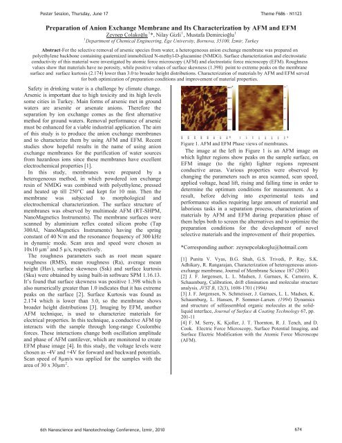
PPoster Session, Thursday, June 17Theme F686 - N1123Preparation of Anion Exchange Membrane and Its Characterization by AFM and EFM111UZeynep ÇolakoluUP P*, Nilay GizliP P, Mustafa DemircioluP1PDepartment of Chemical Eng<strong>in</strong>eer<strong>in</strong>g, Ege University, Bornova, 35100, zmir, TurkeyAbstract-For the selective removal of arsenic species from water, a heterogeneous anion exchange membrane was prepared onpolyethylene backbone conta<strong>in</strong><strong>in</strong>g quaternized immobilized N-methyl-D-glucam<strong>in</strong>e (NMDG). Surface characterization and electrostaticconductivity of this material were <strong>in</strong>vestigated by atomic force microscopy (AFM) and electrostatic force microscopy (EFM). Roughnessvalues show that materials have no porosity, while positive values of surface skewness (1.398) po<strong>in</strong>t to extreme peaks on the membranesurface and surface kurtosis (2.174) lower than 3.0 to broader height distributions. Characterization of materials by AFM and EFM servedfor both optimization of preparation conditions and improvement of material properties.Safety <strong>in</strong> dr<strong>in</strong>k<strong>in</strong>g water is a challenge by climate change.Arsenic is important due to high toxicity and its high levelssome cities <strong>in</strong> Turkey. Ma<strong>in</strong> forms of arsenic met <strong>in</strong> groundwaters are arsenite or arsenate anions. Therefore theseparation by ion exchange comes as the first alternativemethod for ground waters. Removal performance of arsenicmust be enhanced for a viable <strong>in</strong>dustrial application. The aimof this study is to produce the anion exchange membranesand to characterize them by us<strong>in</strong>g AFM and EFM. Recentstudies show hopeful results <strong>in</strong> the name of us<strong>in</strong>g anionexchange membranes for the purification of water sourcesfrom hazardous ions s<strong>in</strong>ce these membranes have excellentelectrochemical properties [1].In this study, membranes were prepared by aheterogeneous method, <strong>in</strong> which powdered ion exchangeres<strong>in</strong> of NMDG was comb<strong>in</strong>ed with polyethylene, pressedand heated up till 250°C and kept for 10 m<strong>in</strong>. Then themembrane was subjected to morphological andelectrochemical characterization. The surface structure ofmembranes was observed by multimode AFM (RT-SHPM,NanoMagnetics Instruments). The membrane surfaces werescanned by alum<strong>in</strong>ium reflex coated silicon probe (Tap300AI, NanoMagnetics Instruments) hav<strong>in</strong>g the spr<strong>in</strong>gconstant of 40 N/m and the resonance frequency of 300 kHz<strong>in</strong> dynamic mode. Scan area and speed were chosen as210x10 μmP Pand 5 μ/s, respectively.The roughness parameters such as root mean squareroughness (RMS), mean roughness (Ra), average meanheight (Hav), surface skewness (Ssk) and surface kurtosis(Sku) were obta<strong>in</strong>ed by us<strong>in</strong>g built-<strong>in</strong> software SPM 1.16.13.It’s found that surface skewness was positive 1.398 which isalso numerically greater than 1.0 <strong>in</strong>dicates that it has extremepeaks on the surface [2]. Surface Kurtosis was found as2.174 which is lower than 3.0, so the membrane showsbroader height distributions [3]. Imag<strong>in</strong>g by EFM, anotherAFM technique, is used to characterize materials forelectrical properties. In this technique, a conductive AFM tip<strong>in</strong>teracts with the sample through long-range Coulombicforces. These <strong>in</strong>teractions change both oscillation amplitudeand phase of AFM cantilever, which are monitored to createEFM phase image [4]. In this study, the voltage levels werechosen as -4V and +4V for forward and backward potentials.Scan speed of 8μm/s was applied for the samples with the2area of 30 x 30μmP P.Figure 1. AFM and EFM Phase views of membranes.The image at the left <strong>in</strong> Figure 1 is an AFM image onwhich lighter regions show peaks on the sample surface, onEFM image (to the right) lighter regions representconductive areas. Various properties were observed bychang<strong>in</strong>g the parameters such as area scanned, scan speed,applied voltage, head lift, ris<strong>in</strong>g and fall<strong>in</strong>g time <strong>in</strong> order todeterm<strong>in</strong>e the optimum conditions for measurement. As aresult, before delv<strong>in</strong>g <strong>in</strong>to experimental tests andperformance studies requir<strong>in</strong>g large amount of material andlaborious tasks <strong>in</strong> a separation process, characterization ofmaterials by AFM and EFM dur<strong>in</strong>g preparation phase ofthem helps both to screen the alternatives and to optimize thepreparation conditions for the development of novelselective materials and the improvement of their properties.*Correspond<strong>in</strong>g author: HTzeynepcolakoglu@hotmail.comT[1] Punita V. Vyas, B.G. Shah, G.S. Trivedi, P. Ray, S.K.Adhikary, R. Rangarajan, Characterization of heterogeneous anionexchangemembrane, Journal of Membrane Science 187 (2001)[2] J. F. Jørgensen, L. L. Madsen, J. Garnaes, K. Carneiro, K.Schaumburg, Calibration, drift elim<strong>in</strong>ation and molecular structureanalysis, JVST B, 12(3), 1698-1701 (1994)[3] J. F. Jørgensen, N. Schmeisser, J. Garnaes, L. L. Madsen, K.Schaumburg, L. Hansen, P. Sommer-Larsen. (1994) Dynamicsand structure of selfassembled organic molecules at the solidliquid<strong>in</strong>terface, Journal of Surface & Coat<strong>in</strong>g Technology 67, pp.201-11[4] F. M. Serry, K. Kjoller, J. T. Thornton, R. J. Tench, and D.Cook. Electric Force Microscopy, Surface Potential Imag<strong>in</strong>g, andSurface Electric Modification with the Atomic Force Microscope(AFM).6th Nanoscience and Nanotechnology Conference, zmir, 2010 674
- Page 1:
Poster Presentations3rd Day17 June
- Page 4 and 5:
Determination of Dielectric Anisotr
- Page 7 and 8:
Poster Session, Thursday, June 17Th
- Page 9 and 10:
PP mPP vs.P =P,PP (1)P andPoster Se
- Page 11 and 12:
PP mPP vs.P =P,PP (1)P andPoster Se
- Page 13 and 14: PP andPoster Session, Thursday, Jun
- Page 15 and 16: Poster Session, Thursday, June 17Th
- Page 17 and 18: PP and770 772 774 776 778 780 782 7
- Page 19 and 20: Poster Session, Thursday, June 17Th
- Page 21 and 22: Poster Session, Thursday, June 17Th
- Page 23 and 24: P25,Poster Session, Thursday, June
- Page 25 and 26: PP TOBBPoster Session, Thursday, Ju
- Page 27 and 28: PisPPisisisP,PisPoster Session, Thu
- Page 29 and 30: U NeslihanPPPPoster Session, Thursd
- Page 31 and 32: Poster Session, Thursday, June 17Th
- Page 33 and 34: PPPoster Session, Thursday, June 17
- Page 35 and 36: PPoster Session, Thursday, June 17T
- Page 37 and 38: P onP viaPP wereP upPoster Session,
- Page 39 and 40: P ·cm.PVPPPsPPPPP andPoster Sessio
- Page 41 and 42: Poster Session, Thursday, June 17Th
- Page 43 and 44: PPoster Session, Thursday, June 17T
- Page 45 and 46: PPoster Session, Thursday, June 17T
- Page 47 and 48: Poster Session, Thursday, June 17Th
- Page 49 and 50: PErkanPoster Session, Thursday, Jun
- Page 51 and 52: Poster Session, Thursday, June 17Th
- Page 53 and 54: Poster Session, Thursday, June 17Th
- Page 55 and 56: PPPP andPoster Session, Thursday, J
- Page 57 and 58: Poster Session, Thursday, June 17Th
- Page 59 and 60: Poster Session, Thursday, June 17Th
- Page 61 and 62: T PeptideTPP,PP,PP andTT2429TTTTTT
- Page 63: Poster Session, Thursday, June 17Th
- Page 67 and 68: Poster Session, Thursday, June 17Th
- Page 69 and 70: PPPoster Session, Thursday, June 17
- Page 71 and 72: Poster Session, Thursday, June 17Th
- Page 73 and 74: Poster Session, Thursday, June 17Th
- Page 75 and 76: PT AdditionalT ThePoster Session, T
- Page 77 and 78: Poster Session, Thursday, June 17Th
- Page 79 and 80: Poster Session, Thursday, June 17Th
- Page 81 and 82: Poster Session, Thursday, June 17Th
- Page 83 and 84: PPoster Session, Thursday, June 17T
- Page 85 and 86: Poster Session, Thursday, June 17Th
- Page 87 and 88: PPPoster Session, Thursday, June 17
- Page 89 and 90: Poster Session, Thursday, June 17Hu
- Page 91 and 92: Poster Session, Thursday, June 17Th
- Page 93 and 94: PPPPPPoster Session, Thursday, June
- Page 95 and 96: Poster Session, Thursday, June 17Th
- Page 97 and 98: Poster Session, Thursday, June 17Th
- Page 99 and 100: Poster Session, Thursday, June 17Th
- Page 101 and 102: PPoster Session, Thursday, June 17T
- Page 103 and 104: Poster Session, Thursday, June 17Th
- Page 105 and 106: PPPPPPPoster Session, Thursday, Jun
- Page 107 and 108: Poster Session, Thursday, June 17Th
- Page 109 and 110: PPPR2R PIN(80)PPgPP OzlemPPoster Se
- Page 111 and 112: Poster Session, Thursday, June 17Th
- Page 113 and 114: Poster Session, Thursday, June 17Th
- Page 115 and 116:
P onPP toP coordinatedPPoster Sessi
- Page 117 and 118:
PPPPP,PP,P(PR RmPoster Session, Thu
- Page 119 and 120:
Poster Session, Thursday, June 17Th
- Page 121 and 122:
Poster Session, Thursday, June 17Th
- Page 123 and 124:
PP InstitutePP DepartmentPoster Ses
- Page 125 and 126:
andPCPPoster Session, Thursday, Jun
- Page 127 and 128:
PP scatteringPYusufPP Corresponding
- Page 129 and 130:
PP toPoster Session, Thursday, June
- Page 131 and 132:
PP andPoster Session, Thursday, Jun
- Page 133 and 134:
PPPPoster Session, Thursday, June 1
- Page 135 and 136:
PPoster Session, Thursday, June 17T
- Page 137 and 138:
PPP andP (.cm).Poster Session, Thur
- Page 139 and 140:
PP tiltP andP editionPoster Session
- Page 141 and 142:
PP andPPoster Session, Thursday, Ju
- Page 143 and 144:
Poster Session, Thursday, June 17Th
- Page 145 and 146:
PP forP forP edit.PPoster Session,
- Page 147 and 148:
Poster Session, Thursday, June 17Th
- Page 149 and 150:
Poster Session, Thursday, June 17Th
- Page 151 and 152:
PP ionicPP ,PPoster Session, Thursd
- Page 153 and 154:
PP lightPoster Session, Thursday, J
- Page 155 and 156:
Poster Session, Thursday, June 17Th
- Page 157 and 158:
PPoster Session, Thursday, June 17T
- Page 159 and 160:
Poster Session, Thursday, June 17Th
- Page 161 and 162:
PandPoster Session, Thursday, June
- Page 163 and 164:
Poster Session, Thursday, June 17 T
- Page 165 and 166:
PPPoster Session, Thursday, June 17
- Page 167 and 168:
PPoster Session, Thursday, June 17T
- Page 169 and 170:
PPoster Session, Thursday, June 17T
- Page 171 and 172:
PPoster Session, Thursday, June 17T
- Page 173 and 174:
PP DepartmentNanoscienceTPPoster Se
- Page 175 and 176:
Poster Session, Thursday, June 17Th
- Page 177 and 178:
Poster Session, Thursday, June 17Th
- Page 179 and 180:
PPPoster Session, Thursday, June 17
- Page 181 and 182:
PPPPPoster Session, Thursday, June
- Page 183 and 184:
PPPPoster Session, Thursday, June 1
- Page 185 and 186:
PPoster Session, Thursday, June 17T
- Page 187 and 188:
PPoster Session, Thursday, June 17T
- Page 189 and 190:
PPoster Session, Thursday, June 17T
- Page 191 and 192:
Poster Session, Thursday, June 17Th
- Page 193 and 194:
Poster Session, Thursday, June 17Th
- Page 195 and 196:
0T0T0T0T AsPPPP werePoster Session,
- Page 197 and 198:
PPoster Session, Thursday, June 17T
- Page 199 and 200:
PPPPPoster Session, Thursday, June
- Page 201 and 202:
PPoster Session, Thursday, June 17T
- Page 203 and 204:
PPoster Session, Thursday, June 17T
- Page 205 and 206:
Poster Session, Thursday, June 17Th
- Page 207 and 208:
PPoster Session, Thursday, June 17T
- Page 209 and 210:
PPoster Session, Thursday, June 17T
- Page 211:
Poster Session, Thursday, June 17AF