PPPPPoster Session, Thursday, June 17Theme F686 - N1123Work Function Difference Measurements of Graphene, Graphite and SiOR2R Surfaces us<strong>in</strong>g Kelv<strong>in</strong>Probe Force Microscope (KPFM)12342UNihan ÖzkanUP P*, Selda SonuenP P, Ümit ÇelikP P, Hidayet Çet<strong>in</strong>P P, Ahmet OralP1PDepartment of Physics Eng<strong>in</strong>eer<strong>in</strong>g, Istanbul Technical University, Istanbul, 34469, Turkey2PFaculty of Eng<strong>in</strong>eer<strong>in</strong>g & Natural Sciences, Sabanci University, Istanbul, 34956, Turkey3PDepartment of Materials Eng<strong>in</strong>eer<strong>in</strong>g, Istanbul Technical University, 34469, Turkey4PDepartment of Physics, Bozok University, Yozgat, TurkeyAbstract-We predict that we can determ<strong>in</strong>e the work function difference between grahene and SiOR2R, graphite and SiOR2and also graphene and graphite. We prepared graphene flakes on SiOR2R substrate by mechanical exfolation method and obta<strong>in</strong>ed images oftopography and work function difference by Kelv<strong>in</strong> Probe Force Microscope.Kelv<strong>in</strong> Probe Force Microscopy (KPFM) is a new localprobe measurement method of work function of differentmetals with high spatial resolution. It depends on theKelv<strong>in</strong> method orig<strong>in</strong>ally developed by William Thomson,also known as Lord Kelv<strong>in</strong> <strong>in</strong> 1898[1]. In 1991 KPFM wasdeveloped by Nonnenmacher et al. to look at differentmetal surfaces with high spatial resolution[2].In this work we modified the Atomic ForceMicroscope(AFM) supplied by NanoMagneticsInstruments Ltd. to perform the Kelv<strong>in</strong> Probe ForceMicroscopy measurements. Graphene flakes were preparedby mechanical exfoliation of graphite <strong>crystals</strong> with tapeand transfer onto Si(100) wafer with 300 nm thermal oxideas shown <strong>in</strong> Figure 1.Figure 2. a)Topography image of multi layer graphene andgraphite by KPFM b)Cross section of topographic KPFM scan ofgrapheneFigure 3. a) Work function difference image of multi layergraphene and graphite by KPFM b) Cross section of workfunction difference scan between graphite and SiOR2R of KPFMFigure 1. Optical microscope image of multi layer grapheneproduced by mechanical exfoliation method, x50 objective lens.There are different KPFM modes commonlyemployed[3] <strong>in</strong> the literature. We operated the KPFM <strong>in</strong>multi-frequency mode <strong>in</strong> this work, topographical signalsand the Kelv<strong>in</strong> probe signal are simultaneously detected atfirst and second resonance frequencies of the cantilever,respectively. Thus topography of the surface and workfunction difference between the sample and tip aresimultaneously determ<strong>in</strong>ed. The first resonance frequencyis used to obta<strong>in</strong> topography image as used <strong>in</strong> <strong>in</strong>termittentcontactAFM mode. The second resonance frequency isused to obta<strong>in</strong> the work function difference image of thesample, us<strong>in</strong>g a digital Lock-<strong>in</strong> Amplifier. An electricalcontact is carefully applied from the side of the flake us<strong>in</strong>gsilver pa<strong>in</strong>t.We obta<strong>in</strong>ed topography images of multilayer and s<strong>in</strong>glelayer graphene as well as graphite flakes deposited onSilicon and measured the thickness of graphene. KPFMimages of the samples were simultaneously obta<strong>in</strong>ed withthe topographic images. We could measure work functiondifference between the graphene, graphite and SiOR2R asshown <strong>in</strong> Figure 2 and Figure 3.RTopography and KPFM images from s<strong>in</strong>gle layergraphene will also be presented [4].This work is supported by TÜBTAK , Project Numbers107T720, 107T892 & 108T930, M<strong>in</strong>istry of Industry &Commerce, Project Number 410.STZ.2009-1 andNanoMagnetics Instruments ltd.*Correspond<strong>in</strong>g author: ozkan.nian@gmail.com[1] Lord Kelv<strong>in</strong>, Contact electricity of metals, Phil. Mag.,46,82(1897)[2 ] Nonnenmacher M., Oboyle M-P.,Wickramas<strong>in</strong>gheH- K., Kelv<strong>in</strong> probe force microscopy, Appl. Phys. Lett.,58 ,2921(1991)[3] Palermo V., Palma M., Samori P.,.Electronic Characterizationof Organic Th<strong>in</strong> Films by Kelv<strong>in</strong> Probe Force Microscopy, Adv.Mater., 18, 145–164(2006)[4] Filleter T., Emtsev K-V., Seyller Th., Bennewitz R.,.LocalWork Function measurements of Epitaxial Graphene, Appl Phys.Lett, 93,133117 (2008)6th Nanoscience and Nanotechnology Conference, zmir, 2010 669
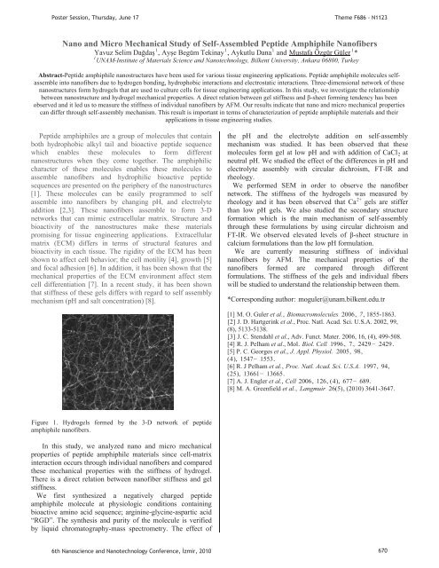
T PeptideTPP,PP,PP andTT2429TTTTTT TTPoster Session, Thursday, June 17Theme F686 - N1123TNano and Micro Mechanical Study of Self-Assembled Peptide Amphiphile Nanofibers1111Yavuz Selim DadaPPAye Begüm Tek<strong>in</strong>ayPPAykutlu DanaPUMustafa Özgür GülerUP P*1PUNAM-Institute of Materials Science and Nanotechnology, Bilkent University, Ankara 06800, TurkeyAbstract-Peptide amphiphile nanostructures have been used for various tissue eng<strong>in</strong>eer<strong>in</strong>g applications. Peptide amphiphile molecules selfassemble<strong>in</strong>to nanofibers due to hydrogen bond<strong>in</strong>g, hydrophobic <strong>in</strong>teractions and electrostatic <strong>in</strong>teractions. Three-dimensional network of thesenanostructures form hydrogels that are used to culture cells for tissue eng<strong>in</strong>eer<strong>in</strong>g applications. In this study, we <strong>in</strong>vestigate the relationshipbetween nanostructure and hydrogel mechanical properties. A direct relation between gel stiffness and -sheet form<strong>in</strong>g tendency has beenobserved and it led us to measure the stiffness of <strong>in</strong>dividual nanofibers by AFM. Our results <strong>in</strong>dicate that nano and micro mechanical propertiescan differ through self-assembly mechanism. This result is important <strong>in</strong> terms of characterization of peptide amphiphile materials and theirapplications <strong>in</strong> tissue eng<strong>in</strong>eer<strong>in</strong>g studies.amphiphiles are a group of molecules that conta<strong>in</strong>both hydrophobic alkyl tail and bioactive peptide sequencewhich enables these molecules to form differentnanostructures when they come together. The amphiphiliccharacter of these molecules enables these molecules toassemble nanofibers and hydrophilic bioactive peptidesequences are presented on the periphery of the nanostructures[1]. These molecules can be easily programmed to selfassemble <strong>in</strong>to nanofibers by chang<strong>in</strong>g pH, and electrolyteaddition [2,3]. These nanofibers assemble to form 3-Dnetworks that can mimic extracellular matrix. Structure andbioactivity of the nanostructures make these materialspromis<strong>in</strong>g for tissue eng<strong>in</strong>eer<strong>in</strong>g applications. Extracellularmatrix (ECM) differs <strong>in</strong> terms of structural features andbioactivity <strong>in</strong> each tissue. The rigidity of the ECM has beenshown to affect cell behavior; the cell motility [4], growth [5]and focal adhesion [6]. In addition, it has been shown that themechanical properties of the ECM environment affect stemcell differentiation [7]. In a recent study, it has been shownthat stiffness of these gels differs with regard to self assemblymechanism (pH and salt concentration) [8].the pH and the electrolyte addition on self-assemblymechanism was studied. It has been observed that thesemolecules form gel at low pH and with addition of CaClR2 Ratneutral pH. We studied the effect of the differences <strong>in</strong> pH andelectrolyte assembly with circular dichroism, FT-IR andrheology.We performed SEM <strong>in</strong> order to observe the nanofibernetwork. The stiffness of the hydrogels was measured by2+rheology and it has been observed that CaPPgels are stifferthan low pH gels. We also studied the secondary structureformation which is the ma<strong>in</strong> mechanism of self-assemblythrough these formulations by us<strong>in</strong>g circular dichroism andFT-IR. We observed elevated levels of -sheet structure <strong>in</strong>calcium formulations than the low pH formulation.We are currently measur<strong>in</strong>g stiffness of <strong>in</strong>dividualnanofibers by AFM. The mechanical properties of thenanofibers formed are compared through differentformulations. The stiffness of the gels and <strong>in</strong>dividual fiberswill be studied to understand the relationship between them.*Correspond<strong>in</strong>g author: HTmoguler@unam.bilkent.edu.trT[1] TM. O. Guler et al., TTBiomacromoleculesTT TT2006T,T TT7T, 1855-1863.T[2]T J. D. Hartger<strong>in</strong>k et al., Proc. Natl. Acad. Sci. U.S.A. 2002, 99,(8), 5133-5138.T[3]T J. C. Stendahl et al., Adv. Funct. Mater. 2006, 16, (4), 499-508.[4] TR. J. Pelham et al., MolTT. Biol. CellTT TT1996TT,TT TT7TT,TT TT2429 TT.[5] P. C. Georges et al., TJ. Appl. Physiol.TT TT2005T,T TT98T,(T4T),T TT1547TT TT1553T.[6] R. J Pelham et al., TProc. Natl. Acad. Sci. U.S.A. 1997T,T TT94T,(T25T),T TT13661TT TT13665T.[7] A. J. Engler et al., TCell TT2006T,T TT126T, (T4T),T TT677TT TT689T.[8] M. A. Greenfield et al.,T TTLangmuirTT TT26(5)T, (2010) 3641-3647.TFigure 1. Hydrogels formed by the 3-D network of peptideamphiphile nanofibers.In this study, we analyzed nano and micro mechanicalproperties of peptide amphiphile materials s<strong>in</strong>ce cell-matrix<strong>in</strong>teraction occurs through <strong>in</strong>dividual nanofibers and comparedthese mechanical properties with the stiffness of hydrogel.There is a direct relation between nanofiber stiffness and gelstiffness. TWe first synthesized a negatively charged peptideamphiphile molecule at physiologic conditions conta<strong>in</strong><strong>in</strong>gbioactive am<strong>in</strong>o acid sequence; arg<strong>in</strong><strong>in</strong>e-glyc<strong>in</strong>e-aspartic acid“RGD”. The synthesis and purity of the molecule is verifiedby liquid chromatography-mass spectrometry. The effect of6th Nanoscience and Nanotechnology Conference, zmir, 2010 670
- Page 1:
Poster Presentations3rd Day17 June
- Page 4 and 5:
Determination of Dielectric Anisotr
- Page 7 and 8:
Poster Session, Thursday, June 17Th
- Page 9 and 10: PP mPP vs.P =P,PP (1)P andPoster Se
- Page 11 and 12: PP mPP vs.P =P,PP (1)P andPoster Se
- Page 13 and 14: PP andPoster Session, Thursday, Jun
- Page 15 and 16: Poster Session, Thursday, June 17Th
- Page 17 and 18: PP and770 772 774 776 778 780 782 7
- Page 19 and 20: Poster Session, Thursday, June 17Th
- Page 21 and 22: Poster Session, Thursday, June 17Th
- Page 23 and 24: P25,Poster Session, Thursday, June
- Page 25 and 26: PP TOBBPoster Session, Thursday, Ju
- Page 27 and 28: PisPPisisisP,PisPoster Session, Thu
- Page 29 and 30: U NeslihanPPPPoster Session, Thursd
- Page 31 and 32: Poster Session, Thursday, June 17Th
- Page 33 and 34: PPPoster Session, Thursday, June 17
- Page 35 and 36: PPoster Session, Thursday, June 17T
- Page 37 and 38: P onP viaPP wereP upPoster Session,
- Page 39 and 40: P ·cm.PVPPPsPPPPP andPoster Sessio
- Page 41 and 42: Poster Session, Thursday, June 17Th
- Page 43 and 44: PPoster Session, Thursday, June 17T
- Page 45 and 46: PPoster Session, Thursday, June 17T
- Page 47 and 48: Poster Session, Thursday, June 17Th
- Page 49 and 50: PErkanPoster Session, Thursday, Jun
- Page 51 and 52: Poster Session, Thursday, June 17Th
- Page 53 and 54: Poster Session, Thursday, June 17Th
- Page 55 and 56: PPPP andPoster Session, Thursday, J
- Page 57 and 58: Poster Session, Thursday, June 17Th
- Page 59: Poster Session, Thursday, June 17Th
- Page 63 and 64: Poster Session, Thursday, June 17Th
- Page 65 and 66: PPoster Session, Thursday, June 17T
- Page 67 and 68: Poster Session, Thursday, June 17Th
- Page 69 and 70: PPPoster Session, Thursday, June 17
- Page 71 and 72: Poster Session, Thursday, June 17Th
- Page 73 and 74: Poster Session, Thursday, June 17Th
- Page 75 and 76: PT AdditionalT ThePoster Session, T
- Page 77 and 78: Poster Session, Thursday, June 17Th
- Page 79 and 80: Poster Session, Thursday, June 17Th
- Page 81 and 82: Poster Session, Thursday, June 17Th
- Page 83 and 84: PPoster Session, Thursday, June 17T
- Page 85 and 86: Poster Session, Thursday, June 17Th
- Page 87 and 88: PPPoster Session, Thursday, June 17
- Page 89 and 90: Poster Session, Thursday, June 17Hu
- Page 91 and 92: Poster Session, Thursday, June 17Th
- Page 93 and 94: PPPPPPoster Session, Thursday, June
- Page 95 and 96: Poster Session, Thursday, June 17Th
- Page 97 and 98: Poster Session, Thursday, June 17Th
- Page 99 and 100: Poster Session, Thursday, June 17Th
- Page 101 and 102: PPoster Session, Thursday, June 17T
- Page 103 and 104: Poster Session, Thursday, June 17Th
- Page 105 and 106: PPPPPPPoster Session, Thursday, Jun
- Page 107 and 108: Poster Session, Thursday, June 17Th
- Page 109 and 110: PPPR2R PIN(80)PPgPP OzlemPPoster Se
- Page 111 and 112:
Poster Session, Thursday, June 17Th
- Page 113 and 114:
Poster Session, Thursday, June 17Th
- Page 115 and 116:
P onPP toP coordinatedPPoster Sessi
- Page 117 and 118:
PPPPP,PP,P(PR RmPoster Session, Thu
- Page 119 and 120:
Poster Session, Thursday, June 17Th
- Page 121 and 122:
Poster Session, Thursday, June 17Th
- Page 123 and 124:
PP InstitutePP DepartmentPoster Ses
- Page 125 and 126:
andPCPPoster Session, Thursday, Jun
- Page 127 and 128:
PP scatteringPYusufPP Corresponding
- Page 129 and 130:
PP toPoster Session, Thursday, June
- Page 131 and 132:
PP andPoster Session, Thursday, Jun
- Page 133 and 134:
PPPPoster Session, Thursday, June 1
- Page 135 and 136:
PPoster Session, Thursday, June 17T
- Page 137 and 138:
PPP andP (.cm).Poster Session, Thur
- Page 139 and 140:
PP tiltP andP editionPoster Session
- Page 141 and 142:
PP andPPoster Session, Thursday, Ju
- Page 143 and 144:
Poster Session, Thursday, June 17Th
- Page 145 and 146:
PP forP forP edit.PPoster Session,
- Page 147 and 148:
Poster Session, Thursday, June 17Th
- Page 149 and 150:
Poster Session, Thursday, June 17Th
- Page 151 and 152:
PP ionicPP ,PPoster Session, Thursd
- Page 153 and 154:
PP lightPoster Session, Thursday, J
- Page 155 and 156:
Poster Session, Thursday, June 17Th
- Page 157 and 158:
PPoster Session, Thursday, June 17T
- Page 159 and 160:
Poster Session, Thursday, June 17Th
- Page 161 and 162:
PandPoster Session, Thursday, June
- Page 163 and 164:
Poster Session, Thursday, June 17 T
- Page 165 and 166:
PPPoster Session, Thursday, June 17
- Page 167 and 168:
PPoster Session, Thursday, June 17T
- Page 169 and 170:
PPoster Session, Thursday, June 17T
- Page 171 and 172:
PPoster Session, Thursday, June 17T
- Page 173 and 174:
PP DepartmentNanoscienceTPPoster Se
- Page 175 and 176:
Poster Session, Thursday, June 17Th
- Page 177 and 178:
Poster Session, Thursday, June 17Th
- Page 179 and 180:
PPPoster Session, Thursday, June 17
- Page 181 and 182:
PPPPPoster Session, Thursday, June
- Page 183 and 184:
PPPPoster Session, Thursday, June 1
- Page 185 and 186:
PPoster Session, Thursday, June 17T
- Page 187 and 188:
PPoster Session, Thursday, June 17T
- Page 189 and 190:
PPoster Session, Thursday, June 17T
- Page 191 and 192:
Poster Session, Thursday, June 17Th
- Page 193 and 194:
Poster Session, Thursday, June 17Th
- Page 195 and 196:
0T0T0T0T AsPPPP werePoster Session,
- Page 197 and 198:
PPoster Session, Thursday, June 17T
- Page 199 and 200:
PPPPPoster Session, Thursday, June
- Page 201 and 202:
PPoster Session, Thursday, June 17T
- Page 203 and 204:
PPoster Session, Thursday, June 17T
- Page 205 and 206:
Poster Session, Thursday, June 17Th
- Page 207 and 208:
PPoster Session, Thursday, June 17T
- Page 209 and 210:
PPoster Session, Thursday, June 17T
- Page 211:
Poster Session, Thursday, June 17AF