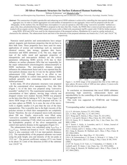
P M.P andP M,Poster Session, Thursday, June 17Theme F686 - N11233D Silver Plasmonic Structure for Surface Enhanced Raman Scatter<strong>in</strong>g11Mehmet KahramanPUMustafa ÇulhaUP P*Genetics and Bioeng<strong>in</strong>eer<strong>in</strong>g Department, Faculty of Eng<strong>in</strong>eer<strong>in</strong>g and Architecture,Yeditepe University, Kayisdagi, Istanbul, TurkeyAbstract- The construction of highly reproducible and enhanc<strong>in</strong>g novel SERS substrate is achieved by controll<strong>in</strong>g the <strong>in</strong>ter-particle distance andaggregate size. In order to control aggregation size and number of nanoparticle <strong>in</strong> one aggregate, micro-well are prepared with the softlithography. In this method, first, the diluted latex microsphere (1.6 μm) are spread as a th<strong>in</strong> film us<strong>in</strong>g “convective assembly” method on aglass slide, then, polydimethylsiloxane (PDMS) is prepared on the latex th<strong>in</strong> film. F<strong>in</strong>ally, the PDMS film is removed and latex particles arewashed with an organic solvent. The PDMS stamp with micro-wells is filled with concentrated AgNPs coated with CTAB for further analysisus<strong>in</strong>g SERS. SEM and AFM were used for the characterization of the prepared surfaces. Rhodam<strong>in</strong>e 6G is used as a probe molecule to7-9characterize the substrate. The enhancement factor and limit of the detection of the prepared substrates are found to be 3.7x10P Pand 1.0x10Prespectively.Nanosize metal particles and semiconductors have uniqueoptical, magnetic and electronic properties that do not have <strong>in</strong>their bulk form. These properties have been used for manyapplications of science and technology such as; nanoscalechemical sensors, data storage, quantum dots lasers,electronics and SERS substrates [1-4]. The size, shape andtype of the noble metal and aggregate properties ofnanoparticles and <strong>in</strong>ter-particle distance are the criticalparameters <strong>in</strong>fluenc<strong>in</strong>g SERS activity [5-8] due to their<strong>in</strong>fluence on surface plasmons (SPs) that are responsible forthe major enhancement (electromagnetic enhancement)[9] <strong>in</strong>SERS mechanism. The <strong>in</strong>ter-particle distance strongly<strong>in</strong>fluences the enhancement factor and it was reported that the<strong>in</strong>ter-particle distance must be 2-4 nm for the optimal SERSenhancement [10]. Although there is an effort to uselithographic methods to control <strong>in</strong>ter-particle distance, thesemethods generally time consum<strong>in</strong>g, expensive and needskilled personnel.In this study, 3D silver plasmonic structures were preparedwith the concentrated silver nanoparticles. First, th<strong>in</strong> film(Figure 1 A) of the latex was prepared us<strong>in</strong>g “convectiveassembly” method [11]. The experimental parameters such asconcentration of latex spheres, mov<strong>in</strong>g stage velocity anddropped volume were studied. Second, PDMS was preparedon the latex th<strong>in</strong> film by bak<strong>in</strong>g at 70 °C for 1 hour. Figure 1 Bshows SEM image of the micro-wells prepared with the 1.6m latex sphere on PDMS. As is seen, the size of the microwellsis slightly smaller (1.4 m) than the size of the latexnanoparticles. This is possibly due to the high viscosity of thepolymer mixture <strong>in</strong> which latex nanoparticles are completelyburied. Therefore, the size of prepared micro-wells decreasesabout 200 nm. F<strong>in</strong>ally, micro-wells were filled us<strong>in</strong>g“convective assembly” method with the concentrated silvernanoparticles conta<strong>in</strong><strong>in</strong>g CTAB, which was used to <strong>in</strong>crease ofthe hydrophobic property of the silver nanoparticles andcontrol <strong>in</strong>ter-particle distance <strong>in</strong> the aggregates (Figure 1C).The enhancement factor was calculated us<strong>in</strong>g IRSERSR/IRBulkR x7CRBulkR/CRSERS Rformula and found as 3.0x10P P. This enhancementfactor is also consistent with 2-4 nm <strong>in</strong>ter-particle distances[12]. The reproducibility of the prepared substrate was testedus<strong>in</strong>g the peak height, area and <strong>in</strong>tensity of the ten peaks at-11512 cmPP. The percent coefficient variance (CV) was foundto be about 10. Limit of the detection (LOD) of the substrate-9was 1.0x10PACFigure 1. A) SEM image of the prepared th<strong>in</strong> film of the 1600 nmlatex sphere, B) micro-wells of 1600 nm latex sphere, C) micro-wellsfilled with silver nanoparticles.In conclusion we demonstrate that novel SERS substratespossess<strong>in</strong>g high sensitivity, enhancement factor andreproducibility by the controll<strong>in</strong>g of the <strong>in</strong>ter-particle distanceand aggregate size.This work was supported by TÜBTAK and YeditepeUniversity.*Correspond<strong>in</strong>g author: HTmculha@yeditepe.edu.trT[1] A. P. Alivisatos, Science 271, 933 (1996).[2] L. E. Brus, Appl. Phys. A, 53, 465 (1991).[3] Y. Wang and N. Herron, J. Phys. Chem. 95, 525 (1991).[4] P. C. Lee and D. Meisel, J. Phys. Chem. 86, 3391 (1982).[5] S. R. Emory, W. E. Hask<strong>in</strong>s, and S. Nie, J. Am. Chem. Soc. 120,8009. (1998).[6] T. Jensen, L. Kelly, A. Lazarides, and G. C. Schatz, Journal ofCluster Science 10, 295 (1999).[7] E. J. Zeman, and G. C. Schatz, J. Phys. Chem. 91, 634 (1987).[8] J. Jiang, K. Bosnick, M. Maillard, and L. Brus, J. Phys. Chem. B107, 9964 (2003).[9] M. Moskovits, Rev. Mod. Phys. 57, 783 (1985).[10] A. M. Schwartzberg, C. D. Grant, A. Wolcott, C. E. Talley, T. R.Huser, R. Bogomolni, and J. Z. Zhang, J. Phys. Chem. B 108, 19191(2004).[11] P. M. Tessier, O. D. Velev, A. T. Kalambur, J. F. Rabolt, A. M.Lenhoff, and E. W. Kaler, J. Am. Chem. Soc. 122, 9554 (2000).[12] J. Jiang, K. Bosnick, M. Maillard, and L. Brus, J. Phys. Chem. B107, 9964 (2003).B6th Nanoscience and Nanotechnology Conference, zmir, 2010 646
P onP viaPP wereP upPoster Session, Thursday, June 17Theme F686 - N1123Superhydrophobic Micropatterned Polymer Surfaces Synthesized by Us<strong>in</strong>g Styrene-Flurometacrylate Random Copolymers11UUur CengizUP P*, H. Yldrm ErbilP1PGebze Institute of Technology, Chemical Eng<strong>in</strong>eer<strong>in</strong>g Department, 41400, Gebze-KocaeliAbstract- In this work, we present a novel method for fabricat<strong>in</strong>g polymer th<strong>in</strong> films conta<strong>in</strong><strong>in</strong>g micro-patterned spherical particles vary<strong>in</strong>g <strong>in</strong>the range of 400 nm to 8 m by dip-coat<strong>in</strong>g process <strong>in</strong> polymer solution. We can control the distribution of the particle size via adjust<strong>in</strong>g theconcentration of the PS-ran-FMA copolymer, the solvent/non-solvent ratio and withdrawal speed of dip coater. Styrene-fluoromethacrylateorandom copolymer were synthesized <strong>in</strong> supercritical carbondioxide (scCOR2R) at 250 bar and 80 P PC us<strong>in</strong>g AIBN as a free radical <strong>in</strong>itiator. It wasfound that the optimal concentration of polymer solution was 25 mg/mL and withdrawal speed of 41 cm/m<strong>in</strong> to obta<strong>in</strong> the narrowest particledistribution on the surface. Surfaces conta<strong>in</strong><strong>in</strong>g the microparticles were characterized with the water contact angle measurement, opticalomicroscopy and SEM. Superhydrophobic surfaces hav<strong>in</strong>g a water contact angle up to 160P obta<strong>in</strong>ed with this novel method.Polymer surfaces composed of two or three dimensionalrepeat<strong>in</strong>g uniform units are called “patterned polymeric1surfaces”P P. These patterned surfaces are referred to micropatternedand nano-patterned surfaces with respect to theirdimensions. The polymeric micro/nano patterns providesome new properties to the surface which change withrespect to chemical nature and shape of the material. For<strong>in</strong>stance, Erbil et al. (2003) obta<strong>in</strong>ed micro-structured gellikeporous super-hydrophobic surfaces hav<strong>in</strong>g a waterocontact angle of 160P the method of phase separationus<strong>in</strong>g isotatic propylene (iPP) hav<strong>in</strong>g a water contact angleoof 105P nonpatterned surfaces with different2solvent/<strong>in</strong>solvent couplesP P. There are other methods toform micro patterned polymeric surfaces. Recently Wanget al. have obta<strong>in</strong>ed micro and nano patterned polymericstructures via phase separation by dropp<strong>in</strong>g polymer1,4solution onto non-solventPP. This method is easier thansoft lithography method whose application is difficult andexpensive.In this study, uniform micro patterned polymericsurfaces were obta<strong>in</strong>ed with particle diameters chang<strong>in</strong>gbetween 400 nm and 8 μm. In the first step, p(ST-ran-FMA) copolymers were synthesized <strong>in</strong> sc-COR2R medium.Styrene and Perfluoromethacrylate (Zonly-TM, Dupont)monomers between 5-20 % <strong>in</strong> molar concentration wereocopolymerized <strong>in</strong> scCOR2 Renvironment at 250 bar and 80P PC.Polymerization <strong>in</strong> COR2 Rhas advantages such as be<strong>in</strong>g nontoxic,cheap and no requirement of extra purificationprocess for the produced copolymers.In the second step, th<strong>in</strong> copolymer film coat<strong>in</strong>gs wereproduced via dip coat<strong>in</strong>g glass slides <strong>in</strong>to polymersolutions obta<strong>in</strong>ed by dissolv<strong>in</strong>g the copolymers <strong>in</strong> THF-MEK mixture (%50 wt) at room temperature and add<strong>in</strong>gmethanol as a non-solvent with vary<strong>in</strong>g amount. Then theoptical and SEM images of the formed surfaces wererecorded and the contact angles of the surface weremeasured by us<strong>in</strong>g the KSV-CAM 200 goniometry.When the methanol volume fraction was low, scatteredform of particles with no specific geometry were observedwhich do not have any specific roughness. With the <strong>in</strong>crease<strong>in</strong> the methanol amount, these particles were converted torepeat<strong>in</strong>g, and somewhat uniform spherical particles. The<strong>in</strong>crease <strong>in</strong> the dipp<strong>in</strong>g rate, the particles shr<strong>in</strong>k uniformly atthe beg<strong>in</strong>n<strong>in</strong>g, but after a certa<strong>in</strong> value of dipp<strong>in</strong>g rate, thenthe agglomeration of particles occurred. Figure 1 shows aSEM image of the surface obta<strong>in</strong>ed at an optimum dipp<strong>in</strong>gspeed and different methanol fraction. It is clearly seen fromthe results particle sizes decrease with the <strong>in</strong>crease ofmethanol fraction. Spherical particles hav<strong>in</strong>g differentdiameters between 2-4 m and 400-800 nm are shown <strong>in</strong>fig.1a and 1b respectivelyFigure 1. SEM images of 25 mg/mL p(ST-ran-FMA) solution <strong>in</strong>THF-MEK solvent mixture (50 wt %) with a) 21,4 b) 33.3 wt %omethanol at 22 P PC mixture temparatureIn summary, particles shape and dimensions and watercontact angle results were varied as a function of nonsolventand copolymer concentration. The <strong>in</strong>crease <strong>in</strong> thenon-solvent fraction resulted <strong>in</strong> decrease of the particlediameter from 8 μm down to 400 nm, and <strong>in</strong>crease <strong>in</strong> theoowater contact angle from 117P to 160P P.* Correspond<strong>in</strong>g author: HTucengiz@gyte.edu.trT[1]Wang Y., Liu Z., Han B., Sun Z., Zhang J., Sun D. Adv.Funct. Mater. 2005, 15, 655.[2]Erbil H.Y., Demirel A.L., Avci Y., Mert O. Science 2003,299, 1377.[3]Xia Y.N., Whitesides G.M., Angew. Chem. Int. Ed. 1998, 37,550.[4] Wang Y., Liu Z.,Huang Y., Han B.,Yang G. Langmuir, 2006,22, 19286th Nanoscience and Nanotechnology Conference, zmir, 2010 647
- Page 1: Poster Presentations3rd Day17 June
- Page 4 and 5: Determination of Dielectric Anisotr
- Page 7 and 8: Poster Session, Thursday, June 17Th
- Page 9 and 10: PP mPP vs.P =P,PP (1)P andPoster Se
- Page 11 and 12: PP mPP vs.P =P,PP (1)P andPoster Se
- Page 13 and 14: PP andPoster Session, Thursday, Jun
- Page 15 and 16: Poster Session, Thursday, June 17Th
- Page 17 and 18: PP and770 772 774 776 778 780 782 7
- Page 19 and 20: Poster Session, Thursday, June 17Th
- Page 21 and 22: Poster Session, Thursday, June 17Th
- Page 23 and 24: P25,Poster Session, Thursday, June
- Page 25 and 26: PP TOBBPoster Session, Thursday, Ju
- Page 27 and 28: PisPPisisisP,PisPoster Session, Thu
- Page 29 and 30: U NeslihanPPPPoster Session, Thursd
- Page 31 and 32: Poster Session, Thursday, June 17Th
- Page 33 and 34: PPPoster Session, Thursday, June 17
- Page 35: PPoster Session, Thursday, June 17T
- Page 39 and 40: P ·cm.PVPPPsPPPPP andPoster Sessio
- Page 41 and 42: Poster Session, Thursday, June 17Th
- Page 43 and 44: PPoster Session, Thursday, June 17T
- Page 45 and 46: PPoster Session, Thursday, June 17T
- Page 47 and 48: Poster Session, Thursday, June 17Th
- Page 49 and 50: PErkanPoster Session, Thursday, Jun
- Page 51 and 52: Poster Session, Thursday, June 17Th
- Page 53 and 54: Poster Session, Thursday, June 17Th
- Page 55 and 56: PPPP andPoster Session, Thursday, J
- Page 57 and 58: Poster Session, Thursday, June 17Th
- Page 59 and 60: Poster Session, Thursday, June 17Th
- Page 61 and 62: T PeptideTPP,PP,PP andTT2429TTTTTT
- Page 63 and 64: Poster Session, Thursday, June 17Th
- Page 65 and 66: PPoster Session, Thursday, June 17T
- Page 67 and 68: Poster Session, Thursday, June 17Th
- Page 69 and 70: PPPoster Session, Thursday, June 17
- Page 71 and 72: Poster Session, Thursday, June 17Th
- Page 73 and 74: Poster Session, Thursday, June 17Th
- Page 75 and 76: PT AdditionalT ThePoster Session, T
- Page 77 and 78: Poster Session, Thursday, June 17Th
- Page 79 and 80: Poster Session, Thursday, June 17Th
- Page 81 and 82: Poster Session, Thursday, June 17Th
- Page 83 and 84: PPoster Session, Thursday, June 17T
- Page 85 and 86: Poster Session, Thursday, June 17Th
- Page 87 and 88:
PPPoster Session, Thursday, June 17
- Page 89 and 90:
Poster Session, Thursday, June 17Hu
- Page 91 and 92:
Poster Session, Thursday, June 17Th
- Page 93 and 94:
PPPPPPoster Session, Thursday, June
- Page 95 and 96:
Poster Session, Thursday, June 17Th
- Page 97 and 98:
Poster Session, Thursday, June 17Th
- Page 99 and 100:
Poster Session, Thursday, June 17Th
- Page 101 and 102:
PPoster Session, Thursday, June 17T
- Page 103 and 104:
Poster Session, Thursday, June 17Th
- Page 105 and 106:
PPPPPPPoster Session, Thursday, Jun
- Page 107 and 108:
Poster Session, Thursday, June 17Th
- Page 109 and 110:
PPPR2R PIN(80)PPgPP OzlemPPoster Se
- Page 111 and 112:
Poster Session, Thursday, June 17Th
- Page 113 and 114:
Poster Session, Thursday, June 17Th
- Page 115 and 116:
P onPP toP coordinatedPPoster Sessi
- Page 117 and 118:
PPPPP,PP,P(PR RmPoster Session, Thu
- Page 119 and 120:
Poster Session, Thursday, June 17Th
- Page 121 and 122:
Poster Session, Thursday, June 17Th
- Page 123 and 124:
PP InstitutePP DepartmentPoster Ses
- Page 125 and 126:
andPCPPoster Session, Thursday, Jun
- Page 127 and 128:
PP scatteringPYusufPP Corresponding
- Page 129 and 130:
PP toPoster Session, Thursday, June
- Page 131 and 132:
PP andPoster Session, Thursday, Jun
- Page 133 and 134:
PPPPoster Session, Thursday, June 1
- Page 135 and 136:
PPoster Session, Thursday, June 17T
- Page 137 and 138:
PPP andP (.cm).Poster Session, Thur
- Page 139 and 140:
PP tiltP andP editionPoster Session
- Page 141 and 142:
PP andPPoster Session, Thursday, Ju
- Page 143 and 144:
Poster Session, Thursday, June 17Th
- Page 145 and 146:
PP forP forP edit.PPoster Session,
- Page 147 and 148:
Poster Session, Thursday, June 17Th
- Page 149 and 150:
Poster Session, Thursday, June 17Th
- Page 151 and 152:
PP ionicPP ,PPoster Session, Thursd
- Page 153 and 154:
PP lightPoster Session, Thursday, J
- Page 155 and 156:
Poster Session, Thursday, June 17Th
- Page 157 and 158:
PPoster Session, Thursday, June 17T
- Page 159 and 160:
Poster Session, Thursday, June 17Th
- Page 161 and 162:
PandPoster Session, Thursday, June
- Page 163 and 164:
Poster Session, Thursday, June 17 T
- Page 165 and 166:
PPPoster Session, Thursday, June 17
- Page 167 and 168:
PPoster Session, Thursday, June 17T
- Page 169 and 170:
PPoster Session, Thursday, June 17T
- Page 171 and 172:
PPoster Session, Thursday, June 17T
- Page 173 and 174:
PP DepartmentNanoscienceTPPoster Se
- Page 175 and 176:
Poster Session, Thursday, June 17Th
- Page 177 and 178:
Poster Session, Thursday, June 17Th
- Page 179 and 180:
PPPoster Session, Thursday, June 17
- Page 181 and 182:
PPPPPoster Session, Thursday, June
- Page 183 and 184:
PPPPoster Session, Thursday, June 1
- Page 185 and 186:
PPoster Session, Thursday, June 17T
- Page 187 and 188:
PPoster Session, Thursday, June 17T
- Page 189 and 190:
PPoster Session, Thursday, June 17T
- Page 191 and 192:
Poster Session, Thursday, June 17Th
- Page 193 and 194:
Poster Session, Thursday, June 17Th
- Page 195 and 196:
0T0T0T0T AsPPPP werePoster Session,
- Page 197 and 198:
PPoster Session, Thursday, June 17T
- Page 199 and 200:
PPPPPoster Session, Thursday, June
- Page 201 and 202:
PPoster Session, Thursday, June 17T
- Page 203 and 204:
PPoster Session, Thursday, June 17T
- Page 205 and 206:
Poster Session, Thursday, June 17Th
- Page 207 and 208:
PPoster Session, Thursday, June 17T
- Page 209 and 210:
PPoster Session, Thursday, June 17T
- Page 211:
Poster Session, Thursday, June 17AF