- Page 2:
Editor Associate Editor for Gynecol
- Page 6:
Editor Dev Maulik, MD, Ph.D. Winthr
- Page 10:
Preface It is with great pleasure w
- Page 16:
Contents 1 Doppler Sonography: A Br
- Page 20:
a Contents XIII Heart and Great Ves
- Page 24:
a Contents XV 15 Pulsed Doppler Ult
- Page 28:
a Contents XVII 22 Doppler Velocime
- Page 32:
a Contents XIX Clinical Application
- Page 36:
a Contents XXI Terbutaline ........
- Page 42:
XXIV Authors Torvid Kiserud, MD, Ph
- Page 46:
2 D. Maulik Fig. 1.2. Title page of
- Page 50:
4 D. Maulik Fig. 1.4. Model of the
- Page 54:
6 D. Maulik Table 1.1. Feasibility
- Page 60:
Chapter 2 Physical Principles of Do
- Page 64:
a Chapter 2 Physical Principles of
- Page 68:
a Chapter 2 Physical Principles of
- Page 72:
a Chapter 2 Physical Principles of
- Page 76:
a Chapter 2 Physical Principles of
- Page 82:
20 D. Maulik one-fourth of a cycle
- Page 86:
22 D. Maulik arterial [6] hemodynam
- Page 90:
24 D. Maulik nomena. The autoregres
- Page 94:
26 D. Maulik far field the beam pro
- Page 98:
28 D. Maulik Mirror Imaging With th
- Page 102:
30 D. Maulik hand, compromise Doppl
- Page 106:
32 D. Maulik vent of broad bandwidt
- Page 110:
34 D. Maulik: Chapter 3 Spectral Do
- Page 114:
36 D. Maulik frequency; the image i
- Page 118:
38 D. Maulik flow directionality du
- Page 122:
40 D. Maulik Doppler Velocity Measu
- Page 126:
42 D. Maulik and for the B-mode ima
- Page 130:
44 D. Maulik known as the Fourier P
- Page 134:
46 D. Maulik Fig. 4.17. Total varia
- Page 138:
48 D. Maulik inverted, but not the
- Page 142:
50 D. Maulik of a ventricular pump,
- Page 146:
52 D. Maulik Table 4.3. Hemodynamic
- Page 150:
54 D. Maulik Placental Vascular Cha
- Page 154:
56 D. Maulik: Chapter 4 Spectral Do
- Page 158:
58 T. Kiserud Fig. 5.2. The impact
- Page 162:
60 T. Kiserud Fig. 5.7. The pressur
- Page 166:
62 T. Kiserud Fig. 5.10. Doppler re
- Page 170:
64 T. Kiserud Fig. 5.13. Upper pane
- Page 174:
66 T. Kiserud of umbilical flow in
- Page 180:
Chapter 6 Sonographic Color Flow Ma
- Page 184:
a Chapter 6 Sonographic Color Flow
- Page 188:
a Chapter 6 Sonographic Color Flow
- Page 192:
a Chapter 6 Sonographic Color Flow
- Page 196:
a Chapter 6 Sonographic Color Flow
- Page 200:
a Chapter 6 Sonographic Color Flow
- Page 204:
a Chapter 6 Sonographic Color Flow
- Page 208:
a Chapter 6 Sonographic Color Flow
- Page 212:
Chapter 7 Three-dimensional Color a
- Page 216:
a Chapter 7 Three-dimensional Color
- Page 220:
a Chapter 7 Three-dimensional Color
- Page 224:
a Chapter 7 Three-dimensional Color
- Page 228:
a Chapter 7 Three-dimensional Color
- Page 232:
Chapter 8 Biological Safety of Diag
- Page 236:
a Chapter 8 Biological Safety of Di
- Page 240:
a Chapter 8 Biological Safety of Di
- Page 244:
a Chapter 8 Biological Safety of Di
- Page 248:
Although the question of inertial o
- Page 252:
a Chapter 8 Biological Safety of Di
- Page 256:
a Chapter 8 Biological Safety of Di
- Page 260:
a Chapter 8 Biological Safety of Di
- Page 264:
a Chapter 8 Biological Safety of Di
- Page 270:
114 J. Itskovitz-Eldor, I. Thaler w
- Page 274:
116 J. Itskovitz-Eldor, I. Thaler d
- Page 278:
118 J. Itskovitz-Eldor, I. Thaler F
- Page 282:
120 J. Itskovitz-Eldor, I. Thaler T
- Page 286:
122 J. Itskovitz-Eldor, I. Thaler t
- Page 290:
124 J. Itskovitz-Eldor, I. Thaler F
- Page 294:
126 J. Itskovitz-Eldor, I. Thaler F
- Page 298:
128 J. Itskovitz-Eldor, I. Thaler 7
- Page 302:
130 J. Itskovitz-Eldor, I. Thaler 8
- Page 308:
Chapter 10 Umbilical Doppler Veloci
- Page 312:
a Chapter 10 Umbilical Doppler Velo
- Page 316:
a Chapter 10 Umbilical Doppler Velo
- Page 320:
a Chapter 10 Umbilical Doppler Velo
- Page 324:
a Chapter 10 Umbilical Doppler Velo
- Page 328:
a Chapter 10 Umbilical Doppler Velo
- Page 334:
146 K. MarsÏ—l Fig. 11.2. Record
- Page 338:
148 K. MarsÏ—l total end-diastol
- Page 342:
150 K. MarsÏ—l Table 11.1. Refer
- Page 346:
152 K. MarsÏ—l Fig. 11.11. Absen
- Page 350:
154 K. MarsÏ—l Diabetes Mellitus
- Page 354:
156 K. MarsÏ—l Table 11.5. Follo
- Page 358:
158 K. MarsÏ—l umbilicalis und d
- Page 364:
Chapter 12 Intrauterine Blood Flow
- Page 368:
a Chapter 12 Intrauterine Blood Flo
- Page 372:
l a Chapter 12 Intrauterine Blood F
- Page 376:
a Chapter 12 Intrauterine Blood Flo
- Page 380:
a Chapter 12 Intrauterine Blood Flo
- Page 384:
a Chapter 12 Intrauterine Blood Flo
- Page 388:
a Chapter 12 Intrauterine Blood Flo
- Page 392:
a Chapter 12 Intrauterine Blood Flo
- Page 398:
178 P. Arbeille et al. Fig. 13.1. I
- Page 402:
180 P. Arbeille et al. Fig. 13.3. I
- Page 406:
182 P. Arbeille et al. Fig. 13.5. a
- Page 410:
184 P. Arbeille et al. Fig. 13.10.
- Page 414:
186 P. Arbeille et al. Cerebral±Um
- Page 418:
188 P. Arbeille et al. moglobin con
- Page 422:
190 P. Arbeille et al. These result
- Page 426:
192 P. Arbeille et al. Fig. 13.15.
- Page 430:
194 P. Arbeille et al. cause IUGR i
- Page 434:
196 P. Arbeille et al. circulation
- Page 440:
Chapter 14 Cerebral Blood Flow Velo
- Page 444:
a Chapter 14 Cerebral Blood Flow Ve
- Page 448:
a Chapter 14 Cerebral Blood Flow Ve
- Page 452:
a Chapter 14 Cerebral Blood Flow Ve
- Page 456:
a Chapter 14 Cerebral Blood Flow Ve
- Page 460:
a Chapter 14 Cerebral Blood Flow Ve
- Page 466:
212 J. C. Veille Doppler Principles
- Page 470:
214 J. C. Veille Fig. 15.5 a±c. No
- Page 474:
216 J. C. Veille Table 15.2. Resist
- Page 478:
218 J. C. Veille response to hypoxi
- Page 482:
220 J. C. Veille Fetal Renal Artery
- Page 486:
222 J. C. Veille cardiac output in
- Page 490:
224 J. C. Veille 28. Silver LE, Des
- Page 496:
Chapter 16 Doppler Velocimetry of t
- Page 500:
a Chapter 16 Doppler Velocimetry of
- Page 504:
a Chapter 16 Doppler Velocimetry of
- Page 508:
a Chapter 16 Doppler Velocimetry of
- Page 512:
a Chapter 16 Doppler Velocimetry of
- Page 516:
a Chapter 16 Doppler Velocimetry of
- Page 520:
a Chapter 16 Doppler Velocimetry of
- Page 524:
a Chapter 16 Doppler Velocimetry of
- Page 528:
a Chapter 16 Doppler Velocimetry of
- Page 532:
a Chapter 16 Doppler Velocimetry of
- Page 536:
a Chapter 16 Doppler Velocimetry of
- Page 540:
a Chapter 16 Doppler Velocimetry of
- Page 544:
a Chapter 16 Doppler Velocimetry of
- Page 548:
a Chapter 16 Doppler Velocimetry of
- Page 552:
Chapter 17 Doppler Velocimetry of t
- Page 556:
a Chapter 17 Doppler Velocimetry of
- Page 560:
data support the view that prostagl
- Page 564:
a Chapter 17 Doppler Velocimetry of
- Page 568:
a Chapter 17 Doppler Velocimetry of
- Page 572:
a Chapter 17 Doppler Velocimetry of
- Page 576:
a Chapter 17 Doppler Velocimetry of
- Page 580:
a Chapter 17 Doppler Velocimetry of
- Page 584:
a Chapter 17 Doppler Velocimetry of
- Page 588:
a Chapter 17 Doppler Velocimetry of
- Page 592:
a Chapter 17 Doppler Velocimetry of
- Page 596:
a Chapter 17 Doppler Velocimetry of
- Page 600:
a Chapter 17 Doppler Velocimetry of
- Page 606:
282 W. J. Ott Table 18.1. Fetal wei
- Page 610:
284 W. J. Ott Fig. 18.4. Umbilical
- Page 614:
286 W. J. Ott diastolic notching in
- Page 618:
288 W. J. Ott Fig. 18.8. Normal (hi
- Page 622:
290 W. J. Ott Fig. 18.10. Normal in
- Page 626:
292 W. J. Ott Fig. 18.14. Non-react
- Page 630:
294 W. J. Ott 21. Tamura RK, Sabbag
- Page 634:
296 W. J. Ott 93. Divon MY (1995) R
- Page 638:
298 W. J. Ott: Chapter 18 Doppler U
- Page 642:
300 H. Odendaal Fig. 19.1. Differen
- Page 646:
302 H. Odendaal Time to Establish t
- Page 650:
304 H. Odendaal were most frequentl
- Page 654:
306 H. Odendaal In addition to lowe
- Page 658:
308 H. Odendaal complications by co
- Page 662:
310 H. Odendaal cal artery: a sign
- Page 668:
Chapter 20 Doppler Velocimetry and
- Page 672:
a Chapter 20 Doppler Velocimetry an
- Page 676:
a Chapter 20 Doppler Velocimetry an
- Page 680:
criterion of a hemoglobin differenc
- Page 684:
a Chapter 20 Doppler Velocimetry an
- Page 688:
a Chapter 20 Doppler Velocimetry an
- Page 692:
a Chapter 20 Doppler Velocimetry an
- Page 696:
a Chapter 20 Doppler Velocimetry an
- Page 700:
a Chapter 20 Doppler Velocimetry an
- Page 704:
Chapter 21 Doppler Sonography in Pr
- Page 708:
a Chapter 21 Doppler Sonography in
- Page 712:
a Chapter 21 Doppler Sonography in
- Page 716:
a Chapter 21 Doppler Sonography in
- Page 722:
340 A. Lysikiewicz [11]. Hypoprotei
- Page 726:
342 A. Lysikiewicz The splenic arte
- Page 730:
344 A. Lysikiewicz Fig. 22.5. Fetal
- Page 734:
346 A. Lysikiewicz Scalp edema and
- Page 738:
348 A. Lysikiewicz selection of the
- Page 742:
350 A. Lysikiewicz References 1. Da
- Page 748:
Chapter 23 Doppler Velocimetry in P
- Page 752:
a Chapter 23 Doppler Velocimetry in
- Page 756:
Redistribution of fetal cardiac out
- Page 760:
a Chapter 23 Doppler Velocimetry in
- Page 764:
a Chapter 23 Doppler Velocimetry in
- Page 768:
Chapter 24 Doppler Velocimetry for
- Page 772:
a Chapter 24 Doppler Velocimetry fo
- Page 776:
a Chapter 24 Doppler Velocimetry fo
- Page 780:
a Chapter 24 Doppler Velocimetry fo
- Page 784:
Although comparisons have been made
- Page 788:
a Chapter 24 Doppler Velocimetry fo
- Page 792:
Chapter 25 Absent End-Diastolic Vel
- Page 796:
a Chapter 25 Absent End-Diastolic V
- Page 800:
a Chapter 25 Absent End-Diastolic V
- Page 804:
a Chapter 25 Absent End-Diastolic V
- Page 808:
The homeostatic significance of abs
- Page 812:
a Chapter 25 Absent End-Diastolic V
- Page 816:
Chapter 26 Doppler Velocimetry for
- Page 820:
a Chapter 26 Doppler Velocimetry fo
- Page 824:
a Chapter 26 Doppler Velocimetry fo
- Page 828:
a Chapter 26 Doppler Velocimetry fo
- Page 832:
a Chapter 26 Doppler Velocimetry fo
- Page 836:
striction or hypertensive disease o
- Page 840:
a Chapter 26 Doppler Velocimetry fo
- Page 844:
a Chapter 26 Doppler Velocimetry fo
- Page 850:
404 Y. Chiba et al. apex at ventric
- Page 854:
406 Y. Chiba et al. Fig. 27.5. An e
- Page 858:
408 Y. Chiba et al. Fig. 27.12. Bip
- Page 862:
410 Y. Chiba et al. Fig. 27.16. Blo
- Page 868:
Chapter 28 Ductus Venosus Torvid Ki
- Page 872:
a Chapter 28 Ductus Venosus 415 low
- Page 876:
a Chapter 28 Ductus Venosus 417 Fig
- Page 880:
a Chapter 28 Ductus Venosus 419 Fig
- Page 884:
a Chapter 28 Ductus Venosus 421 mor
- Page 888:
a Chapter 28 Ductus Venosus 423 sli
- Page 892:
a Chapter 28 Ductus Venosus 425 men
- Page 896:
a Chapter 28 Ductus Venosus 427 119
- Page 902:
430 A.A. Baschat coronary vessels a
- Page 906:
432 A.A. Baschat Fig. 29.2. The ori
- Page 910:
434 A.A. Baschat Fig. 29.6. The fet
- Page 914:
436 A.A. Baschat Clinical Applicati
- Page 918:
438 A.A. Baschat agement [67±69].
- Page 922:
440 A.A. Baschat 20. Spahr R, Probs
- Page 928:
Chapter 30 Doppler Interrogation of
- Page 932:
a Chapter 30 Doppler Interrogation
- Page 936:
a Chapter 30 Doppler Interrogation
- Page 940:
a Chapter 30 Doppler Interrogation
- Page 946:
452 R. Chaoui et al. Fig. 31.2. Api
- Page 950:
454 R. Chaoui et al. Fig. 31.7. Spe
- Page 954:
456 R. Chaoui et al. supported by a
- Page 958:
458 R. Chaoui et al. Anomalies of t
- Page 962:
460 R. Chaoui et al. Fig. 31.19. Ca
- Page 966:
462 R. Chaoui et al. Fig. 31.23. In
- Page 972:
Chapter 32 Introduction to Fetal Do
- Page 976:
a Chapter 32 Introduction to Fetal
- Page 980:
a Chapter 32 Introduction to Fetal
- Page 984:
a Chapter 32 Introduction to Fetal
- Page 988:
a Chapter 32 Introduction to Fetal
- Page 992:
a Chapter 32 Introduction to Fetal
- Page 996: a Chapter 32 Introduction to Fetal
- Page 1000: a Chapter 32 Introduction to Fetal
- Page 1004: a Chapter 32 Introduction to Fetal
- Page 1008: a Chapter 32 Introduction to Fetal
- Page 1014: 486 D. Maulik Table 33.2. Approxima
- Page 1018: 488 D. Maulik report in utero worse
- Page 1022: 490 D. Maulik Fig. 33.3. Spectral D
- Page 1026: 492 D. Maulik Fig. 33.7. Color Dopp
- Page 1030: 494 D. Maulik Fig. 33.11. Color flo
- Page 1034: 496 D. Maulik Fig. 33.14 A,B. Fetal
- Page 1038: 498 D. Maulik anteroposterior devia
- Page 1042: 500 D. Maulik Fig. 33.20. Echocardi
- Page 1046: 502 D. Maulik Fig. 33.23. Pulmonary
- Page 1052: a Chapter 33 Doppler Echocardiograp
- Page 1056: a Chapter 33 Doppler Echocardiograp
- Page 1062: 510 D. Maulik true real-time 4D ech
- Page 1066: 512 D. Maulik Fig. 34.6. Two-dimens
- Page 1070: 514 D. Maulik Fig. 34.11. Four-dime
- Page 1074: 516 D. Maulik: Chapter 34 Four-Dime
- Page 1078: 518 W. J. Ott Fig. 35.2. The oxygen
- Page 1082: 520 W. J. Ott Table 35.1. Causes of
- Page 1086: 522 W. J. Ott Velocity Measurements
- Page 1090: 524 W. J. Ott Fig. 35.9. Doppler bl
- Page 1094: 526 W. J. Ott 5. The presence of va
- Page 1098:
528 W. J. Ott Fig. 35.10. Regressio
- Page 1102:
530 W. J. Ott Fig. 35.11. The signi
- Page 1106:
532 W. J. Ott walls in fetuses of d
- Page 1110:
534 W. J. Ott 39. Sharf M, Abinader
- Page 1114:
536 W. J. Ott: Chapter 35 Doppler E
- Page 1118:
538 D. Arduini, G. Rizzo General Pr
- Page 1122:
540 D. Arduini, G. Rizzo tween the
- Page 1126:
542 D. Arduini, G. Rizzo Fig. 36.2.
- Page 1130:
544 D. Arduini, G. Rizzo the condit
- Page 1134:
546 D. Arduini, G. Rizzo: Chapter 3
- Page 1138:
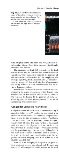
548 J. C. Huhta et al. The equipmen
- Page 1142:
550 J. C. Huhta et al. presence of
- Page 1146:
552 J. C. Huhta et al. say of the i
- Page 1150:
554 J. C. Huhta et al. amination. I
- Page 1154:
556 J. C. Huhta et al.: Chapter 37
- Page 1158:
558 I. Zalud, L. D. Platt Fig. 38.1
- Page 1162:
560 I. Zalud, L. D. Platt Fig. 38.3
- Page 1166:
562 I. Zalud, L. D. Platt were non-
- Page 1170:
564 I. Zalud, L. D. Platt two benig
- Page 1174:
566 I. Zalud, L. D. Platt Fig. 38.6
- Page 1178:
568 I. Zalud, L. D. Platt: Chapter
- Page 1182:
570 I. Zalud and pulsed-wave Dopple
- Page 1186:
572 I. Zalud Fig. 39.4. Corpus lute
- Page 1190:
574 I. Zalud Table 39.2. Data on wo
- Page 1194:
576 I. Zalud corpus luteum, respect
- Page 1198:
578 I. Zalud Previous studies have
- Page 1202:
580 I. Zalud cavity is ªcoldº wit
- Page 1206:
582 I. Zalud Fig. 39.20. Ipsilatera
- Page 1210:
584 I. Zalud However, recent advanc
- Page 1214:
586 I. Zalud [60]. If a 50-mg dose
- Page 1218:
588 I. Zalud ous increase in the RI
- Page 1222:
590 I. Zalud production of progeste
- Page 1226:
592 I. Zalud Fig. 39.33. Pulsed-wav
- Page 1230:
594 I. Zalud Fig. 39.37. Luteal blo
- Page 1234:
596 I. Zalud cal aspects and diagno
- Page 1240:
Chapter 40 Doppler Ultrasonography
- Page 1244:
a Chapter 40 Doppler Ultrasonograph
- Page 1248:
a Chapter 40 Doppler Ultrasonograph
- Page 1252:
a Chapter 40 Doppler Ultrasonograph
- Page 1256:
a Chapter 40 Doppler Ultrasonograph
- Page 1260:
a Chapter 40 Doppler Ultrasonograph
- Page 1266:
612 Subject Index anomalous ± A-V
- Page 1270:
614 Subject Index ± ± cerebral va
- Page 1274:
616 Subject Index ± ± absence of
- Page 1278:
618 Subject Index ± cerebral Doppl
- Page 1282:
620 Subject Index ± therapy 553 in
- Page 1286:
622 Subject Index ± systolic/diast
- Page 1290:
624 Subject Index postextrasystolic
- Page 1294:
626 Subject Index serum estradiol c
- Page 1298:
628 Subject Index trisomy 525 troph
- Page 1302:
630 Subject Index visceral ± malro


