Datascope Passport - Mindray
Datascope Passport - Mindray
Datascope Passport - Mindray

You also want an ePaper? Increase the reach of your titles
YUMPU automatically turns print PDFs into web optimized ePapers that Google loves.
Operation Arrhythmia Algorithm (Optional <strong>Passport</strong> 2)<br />
Heart Rate Meter<br />
Heart Rate is computed using the 16 most recent R-R intervals for heart rates above 48 beats<br />
per minute. If the rate calculated using the last 4 beats is less than 48 beats per minute, then<br />
this rate is used. All detected beats are used to compute the heart rate. A separate ventricular<br />
rate is used in the algorithm to determine rhythms like ventricular tachycardia and ventricular<br />
run.<br />
Filtering Pacer Signals<br />
In order to prevent pacer pulses from being mistaken for QRS complexes, they are removed<br />
from the ECG data that is sent to the arrhythmia algorithm for analysis. Pacer pulses are<br />
shown on the <strong>Passport</strong> 2 as exaggerated vertical lines.<br />
ECG Amplitude<br />
The QRS detection threshold algorithm setting is fixed between 0.15 and 0.45 mV to avoid<br />
detecting noise spikes or P-waves as valid beats. Changing the display gain on the monitor<br />
does not affect the signal that is used by the algorithm for beat detection. For optimal<br />
performance, the leads selected for monitoring should have an amplitude of 0.5 to 1 mV or<br />
more.<br />
Learning<br />
The process of learning is used to establish a normal beat template for a patient. The learn<br />
period is dependent on the heart rate and the dominant pattern. Learning should not be<br />
initiated during a primarily ventricular rhythm because an ectopic beat may be established<br />
as normal.<br />
A learn should be initiated when beats are not being properly detected, or when they are<br />
being erroneously classified. However, if a signal is not strong enough, or lead data is<br />
extremely noisy, better signal quality must be established before a learn can be effective.<br />
Beat Detection and Typing<br />
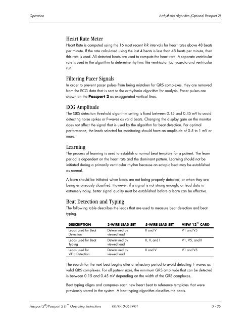
The following table describes the leads that are used to measure beat detection and beat<br />
typing.<br />
DESCRIPTION 3-WIRE LEAD SET 5-WIRE LEAD SET VIEW 12 CARD<br />
Leads used for Beat<br />
Detection<br />
Leads used for Beat<br />
Typing<br />
Leads used for<br />
V-Fib Detection<br />
Determined by<br />
viewed lead<br />
Determined by<br />
viewed lead<br />
Determined by<br />
viewed lead<br />
II and V V1 and V5<br />
II, V, and I V1, V5, and II<br />
II and V V1 and V5<br />
The search for the next beat begins after a refractory period to avoid detecting T- waves as<br />
valid QRS complexes. For all patient sizes, the minimum QRS amplitude that can be detected<br />
is between 0.15 and 0.45 mV depending on the width of the QRS complexes.<br />
Beat typing aligns and compares each new heart beat to reference templates that were<br />
previously stored in the system. A beat typing algorithm classifies the beats.<br />
<strong>Passport</strong> 2 ® /<strong>Passport</strong> 2 LT Operating Instructions 0070-10-0649-01 3 - 35