The Influence Of Priming Two Cucumber Cultivar Seeds
The Influence Of Priming Two Cucumber Cultivar Seeds
The Influence Of Priming Two Cucumber Cultivar Seeds

Create successful ePaper yourself
Turn your PDF publications into a flip-book with our unique Google optimized e-Paper software.
J. Duhok Univ. Vol.13, No.1, (Agri. And Vet. Sciences) Pp 153-161, 2010<br />
from local breed of black goats from 35 flocks of<br />
different localities (districts and sub districts)<br />
which include Zakho, Batel, Sumaill, Aqra and<br />
Center of Duhok province.<br />
2. Blood Smears<br />
Method of preparation of blood smears and<br />
Giemsa staining was used as described by Adam<br />
et al., (1971). <strong>The</strong> thin and thick blood smears<br />
were prepared from the peripheral ear vein.<br />
Before the sample, the site was punctured,<br />
clipped and wiped with 70% ethanol and<br />
allowed to dry the first drop of blood always<br />
discarded, a drop of blood was taken on a clean<br />
glass microscope slide, spread by another slide<br />
at an acute angle; air dried, and fixed by<br />
methanol 5 minute and stained with 10% Giemsa<br />
stain. 30 minutes<br />
3. Serologic Test for Detection of A. ovis<br />
<strong>The</strong> anaplasmosis competitive enzyme-linked<br />
immunosorbent assay (cELISA) was performed<br />
with serum sample using the Anaplasma<br />
Antibody Test Kit, cELISA from VMRD<br />
Inc.(2006) (Pullman, WA, USA) (Knowles et<br />
al., 1996) following the manufactures’<br />
instructions. This assay detects serum antibodies<br />
to a major surface protein (MSP5) of A.<br />
marginale, A. centrale, A. ovis and A.<br />
phagocytophilum (Dreher et al., 2005).<br />
4. Hematological Examination:<br />
Ten milliliters of blood was drained from<br />
jugular vein puncture, after the area was<br />
prepared aseptically, by clipping and then<br />
154<br />
soaking with 70% ethyl alcohol. Three milliliters<br />
of whole blood were mixed in disposable clean<br />
plastic tube with an anticoagulant (EDTA) and<br />
used for the estimation hematological<br />
parameters, while remaining seven milliliters of<br />
the blood were deposited in tube free from<br />
anticoagulant, to obtain serum for estimation of<br />
serological test by ordinary centrifuge 5000 rpm<br />
spanned for 5 minutes and separated serum kept<br />
at – 20°C until used. Hematological parameters<br />
included, total erythrocyte count (RBC), packed<br />
cell volume (PCV), hemoglobin concentration<br />
(Hb), differential leukocyte count, mean<br />
corpuscular volume (MCV), mean corpuscular<br />
hemoglobin (MCH), and mean corpuscular<br />
hemoglobin concentration (MCHC), were done<br />
by methods of Coles (1986).<br />
Statistical analysis:<br />
<strong>The</strong> results were analyzed by chi- square test<br />
(X 2 ) (Tekin, 2003).<br />
RESULTS<br />
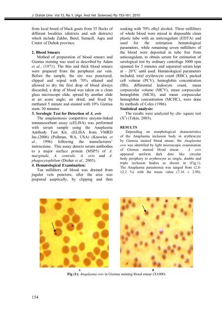
Depending on morphological characteristics<br />
of the Anaplasma inclusion body in erythrocyte<br />
by Giemsa stained blood smear, the Anaplasma<br />
ovis was identified by light microscopic examination<br />
of Giemsa stained blood smear. A. ovis<br />
appeared uniform dark dote like circular<br />
body periphery to erythrocyte as single, double and<br />
triple inclusion bodies as shown in (Fig.1).<br />
<strong>The</strong> Anaplasma parasitemia was ranged from (2.8-<br />
12.2 %) with the mean value (7.16 ± 2.98).<br />
A B<br />
Fig (1): Anaplasma ovis in Giemsa staining blood smear (X1000)