

Monograph on the Potential Human Reproductive and ... - OEHHA
Monograph on the Potential Human Reproductive and ... - OEHHA
Monograph on the Potential Human Reproductive and ... - OEHHA

Create successful ePaper yourself
Turn your PDF publications into a flip-book with our unique Google optimized e-Paper software.
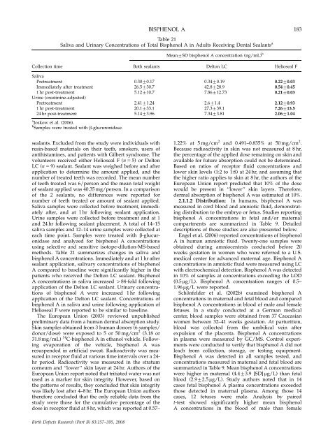
BISPHENOL A<br />
Table 21<br />
Saliva <strong>and</strong> Urinary C<strong>on</strong>centrati<strong>on</strong>s of Total Bisphenol A in Adults Receiving Dental Sealants a<br />
Mean7SD bisphenol A c<strong>on</strong>centrati<strong>on</strong> (ng/mL) b<br />
Collecti<strong>on</strong> time Both sealants Delt<strong>on</strong> LC Helioseal F<br />
Saliva<br />
Pretreatment 0.3070.17 0.3470.19 0.2270.03<br />
Immediately after treatment 26.5730.7 42.8728.9 0.5470.45<br />
1 hr post-treatment 5.12710.7 7.86712.73 0.2170.03<br />
Urine (creatinine-adjusted)<br />
Pretreatment 2.4171.24 2.671.4 2.1270.93<br />
1 hr post-treatment 20.1733.1 27.3739.1 7.26713.5<br />
24 hr post-treatment 5.1473.96 7.3473.81 2.0671.04<br />
a Joskow et al. (2006).<br />
b Samples were treated with b-glucur<strong>on</strong>idase.<br />
sealants. Excluded from <strong>the</strong> study were individuals with<br />
resin-based materials <strong>on</strong> <strong>the</strong>ir teeth, smokers, users of<br />
antihistamines, <strong>and</strong> patients with Gilbert syndrome. The<br />
volunteers received ei<strong>the</strong>r Helioseal F (n 5 5) or Delt<strong>on</strong><br />
LC (n 5 9) sealant. Sealant was weighed before <strong>and</strong> after<br />
applicati<strong>on</strong> to determine <strong>the</strong> amount applied, <strong>and</strong> <strong>the</strong><br />
number of treated teeth was recorded. The mean number<br />
of teeth treated was 6/pers<strong>on</strong> <strong>and</strong> <strong>the</strong> mean total weight<br />
of sealant applied was 40.35 mg/pers<strong>on</strong>. In a comparis<strong>on</strong><br />
of <strong>the</strong> 2 sealants, no differences were reported for<br />
number of teeth treated or amount of sealant applied.<br />
Saliva samples were collected before treatment, immediately<br />
after, <strong>and</strong> at 1 hr following sealant applicati<strong>on</strong>.<br />
Urine samples were collected before treatment <strong>and</strong> at 1<br />
<strong>and</strong> 24 hr following sealant placement. A total of 14–15<br />
saliva samples <strong>and</strong> 12–14 urine samples were collected at<br />
each time point. Samples were treated with b-glucur<strong>on</strong>idase<br />
<strong>and</strong> analyzed for bisphenol A c<strong>on</strong>centrati<strong>on</strong>s<br />
using selective <strong>and</strong> sensitive isotope-diluti<strong>on</strong>-MS-based<br />
methods. Table 21 summarizes changes in saliva <strong>and</strong><br />
bisphenol A c<strong>on</strong>centrati<strong>on</strong>s. Immediately <strong>and</strong> at 1 hr after<br />
sealant applicati<strong>on</strong>, salivary c<strong>on</strong>centrati<strong>on</strong>s of bisphenol<br />
A compared to baseline were significantly higher in <strong>the</strong><br />
patients who received <strong>the</strong> Delt<strong>on</strong> LC sealant. Bisphenol<br />
A c<strong>on</strong>centrati<strong>on</strong>s in saliva increased 484-fold following<br />
applicati<strong>on</strong> of <strong>the</strong> Delt<strong>on</strong> LC sealant. Urinary c<strong>on</strong>centrati<strong>on</strong>s<br />
of bisphenol A were increased 1 hr following<br />
applicati<strong>on</strong> of <strong>the</strong> Delt<strong>on</strong> LC sealant. C<strong>on</strong>centrati<strong>on</strong>s of<br />
bisphenol A in saliva <strong>and</strong> urine following applicati<strong>on</strong> of<br />
Helioseal F were reported to be similar to baseline.<br />
The European Uni<strong>on</strong> (2003) reviewed unpublished<br />
preliminary data from a human dermal absorpti<strong>on</strong> study.<br />
Skin samples obtained from 3 human d<strong>on</strong>ors (6 samples/<br />
d<strong>on</strong>or/dose) were exposed to 5 or 50 mg/cm 2 (3.18 or<br />
31.8 mg/mL) 14 C-bisphenol A in ethanol vehicle. Following<br />
evaporati<strong>on</strong> of <strong>the</strong> vehicle, bisphenol A was<br />
resuspended in artificial sweat. Radioactivity was measured<br />
in receptor fluid at various time intervals over a 24hr<br />
period. Radioactivity was measured in <strong>the</strong> stratum<br />
corneum <strong>and</strong> ‘‘lower’’ skin layer at 24 hr. Authors of <strong>the</strong><br />
European Uni<strong>on</strong> report noted that tritiated water was not<br />
used as a marker for skin integrity. However, based <strong>on</strong><br />
<strong>the</strong> patterns of results, <strong>the</strong>y c<strong>on</strong>cluded that skin integrity<br />
was likely lost after 4–8 hr. The European Uni<strong>on</strong> authors<br />
<strong>the</strong>refore c<strong>on</strong>cluded that <strong>the</strong> <strong>on</strong>ly reliable data from <strong>the</strong><br />
study were those for <strong>the</strong> cumulative percentage of <strong>the</strong><br />
dose in receptor fluid at 8 hr, which was reported at 0.57–<br />
Birth Defects Research (Part B) 83:157–395, 2008<br />
183<br />
1.22% at 5 mg/cm 2 <strong>and</strong> 0.491–0.835% at 50 mg/cm 2 .<br />
Because radioactivity in skin was not measured at 8 hr,<br />
<strong>the</strong> percentage of <strong>the</strong> applied dose remaining <strong>on</strong> skin <strong>and</strong><br />
available for future absorpti<strong>on</strong> could not be determined.<br />
Based <strong>on</strong> ratios of receptor fluid c<strong>on</strong>centrati<strong>on</strong>s <strong>and</strong><br />
lower skin levels (1:2 to 1:8) at 24 hr, <strong>and</strong> assuming that<br />
<strong>the</strong> higher ratio applies to skin at 8 hr, <strong>the</strong> authors of <strong>the</strong><br />
European Uni<strong>on</strong> report predicted that 10% of <strong>the</strong> dose<br />
would be present in ‘‘lower’’ skin layers. Therefore,<br />
dermal absorpti<strong>on</strong> of bisphenol A was estimated at 10%.<br />
2.1.1.2 Distributi<strong>on</strong>: In humans, bisphenol A was<br />
measured in cord blood <strong>and</strong> amniotic fluid, dem<strong>on</strong>strating<br />
distributi<strong>on</strong> to <strong>the</strong> embryo or fetus. Studies reporting<br />
bisphenol A c<strong>on</strong>centrati<strong>on</strong>s in fetal <strong>and</strong>/or maternal<br />
compartments are summarized in Table 9. Detailed<br />
descripti<strong>on</strong>s of those studies are also presented below.<br />
Engel et al. (2006) reported c<strong>on</strong>centrati<strong>on</strong>s of bisphenol<br />
A in human amniotic fluid. Twenty-<strong>on</strong>e samples were<br />
obtained during amniocentesis c<strong>on</strong>ducted before 20<br />
weeks gestati<strong>on</strong> in women who were referred to a U.S.<br />
medical center for advanced maternal age. Bisphenol A<br />
c<strong>on</strong>centrati<strong>on</strong>s in amniotic fluid were measured using LC<br />
with electrochemical detecti<strong>on</strong>. Bisphenol A was detected<br />
in 10% of samples at c<strong>on</strong>centrati<strong>on</strong>s exceeding <strong>the</strong> LOD<br />
(0.5 mg/L). Bisphenol A c<strong>on</strong>centrati<strong>on</strong> ranges of 0.5–<br />
1.96 mg/L were reported.<br />
Schönfelder et al. (2002b) examined bisphenol A<br />
c<strong>on</strong>centrati<strong>on</strong>s in maternal <strong>and</strong> fetal blood <strong>and</strong> compared<br />
bisphenol A c<strong>on</strong>centrati<strong>on</strong>s in blood of male <strong>and</strong> female<br />
fetuses. In a study c<strong>on</strong>ducted at a German medical<br />
center, blood samples were obtained from 37 Caucasian<br />
women between 32–41 weeks gestati<strong>on</strong>. At parturiti<strong>on</strong>,<br />
blood was collected from <strong>the</strong> umbilical vein after<br />
expulsi<strong>on</strong> of <strong>the</strong> placenta. Bisphenol A c<strong>on</strong>centrati<strong>on</strong>s<br />
in plasma were measured by GC/MS. C<strong>on</strong>trol experiments<br />
were c<strong>on</strong>ducted to verify that bisphenol A did not<br />
leach from collecti<strong>on</strong>, storage, or testing equipment.<br />
Bisphenol A was detected in all samples tested, <strong>and</strong><br />
c<strong>on</strong>centrati<strong>on</strong>s measured in maternal <strong>and</strong> fetal blood are<br />
summarized in Table 9. Mean bisphenol A c<strong>on</strong>centrati<strong>on</strong>s<br />
were higher in maternal (4.473.9 [SD] mg/L) than fetal<br />
blood (2.972.5 mg/L). Study authors noted that in 14<br />
cases fetal bisphenol A plasma c<strong>on</strong>centrati<strong>on</strong>s exceeded<br />
those detected in maternal plasma. Am<strong>on</strong>g those 14<br />
cases, 12 fetuses were male. Analysis by paired<br />
t-test showed significantly higher mean bisphenol<br />
A c<strong>on</strong>centrati<strong>on</strong>s in <strong>the</strong> blood of male than female


