

Monograph on the Potential Human Reproductive and ... - OEHHA
Monograph on the Potential Human Reproductive and ... - OEHHA
Monograph on the Potential Human Reproductive and ... - OEHHA

Create successful ePaper yourself
Turn your PDF publications into a flip-book with our unique Google optimized e-Paper software.
in each group were killed at 96 hr post-dosing. Maternal<br />
organs, 6 embryos or fetuses/dam (when possible), <strong>and</strong><br />
placentas were collected. Samples were analyzed for<br />
radioactivity <strong>and</strong> bisphenol A <strong>and</strong>/or bisphenol A<br />
glucur<strong>on</strong>ide by HPLC/liquid scintillati<strong>on</strong> spectrometry.<br />
In all groups, 90–94% of radioactivity was recovered.<br />
Eliminati<strong>on</strong> of bisphenol A <strong>and</strong> its metabolites is<br />
discussed in Secti<strong>on</strong> 2.1.2.4. At 96 hr following dosing,<br />
low percentages of <strong>the</strong> dose were present in carcass (B1–<br />
6%) <strong>and</strong> tissues such as brain, fat, liver, kidney, ovary,<br />
uterus, <strong>and</strong> skin. The <strong>on</strong>ly quantifiable data in placentas<br />
<strong>and</strong> fetuses at 96 hr were obtained in <strong>the</strong> GD 17 group,<br />
<strong>and</strong> those samples c<strong>on</strong>tained 0.01–0.07% of <strong>the</strong> bisphenol<br />
A dose. St<strong>and</strong>ard deviati<strong>on</strong>s for maternal <strong>and</strong> fetal<br />
tissues generally exceeded 50% of <strong>the</strong> mean. Study<br />
authors c<strong>on</strong>cluded that dispositi<strong>on</strong> of radioactivity was<br />
similar in pregnant <strong>and</strong> n<strong>on</strong>-pregnant rats.<br />
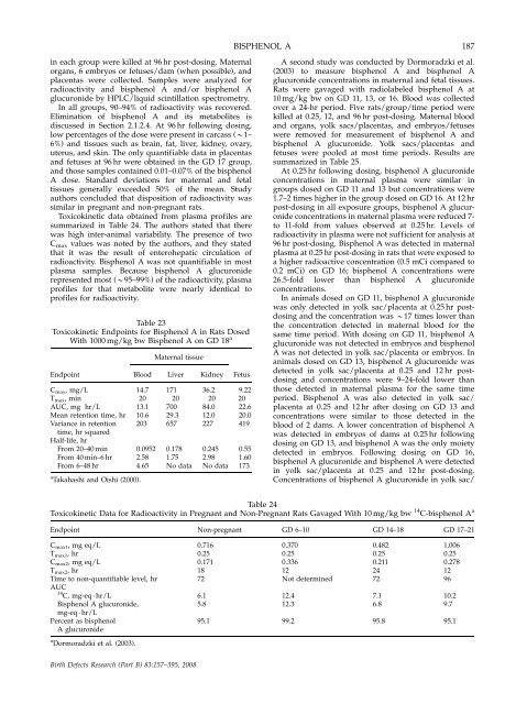
Toxicokinetic data obtained from plasma profiles are<br />
summarized in Table 24. The authors stated that <strong>the</strong>re<br />
was high inter-animal variability. The presence of two<br />
C max values was noted by <strong>the</strong> authors, <strong>and</strong> <strong>the</strong>y stated<br />
that it was <strong>the</strong> result of enterohepatic circulati<strong>on</strong> of<br />
radioactivity. Bisphenol A was not quantifiable in most<br />
plasma samples. Because bisphenol A glucur<strong>on</strong>ide<br />
represented most (B95–99%) of <strong>the</strong> radioactivity, plasma<br />
profiles for that metabolite were nearly identical to<br />
profiles for radioactivity.<br />
Table 23<br />
Toxicokinetic Endpoints for Bisphenol A in Rats Dosed<br />
With 1000 mg/kg bw Bisphenol A <strong>on</strong> GD 18 a<br />
Maternal tissue<br />
Endpoint Blood Liver Kidney Fetus<br />
C max, mg/L 14.7 171 36.2 9.22<br />
T max, min 20 20 20 20<br />
AUC, mg hr/L 13.1 700 84.0 22.6<br />
Mean retenti<strong>on</strong> time, hr 10.6 29.3 12.0 20.0<br />
Variance in retenti<strong>on</strong> 203 657 227 419<br />
time, hr squared<br />
Half-life, hr<br />
From 20–40 min 0.0952 0.178 0.245 0.55<br />
From 40 min–6 hr 2.58 1.75 2.98 1.60<br />
From 6–48 hr 4.65 No data No data 173<br />
a Takahashi <strong>and</strong> Oishi (2000).<br />
BISPHENOL A<br />
187<br />
A sec<strong>on</strong>d study was c<strong>on</strong>ducted by Dormoradzki et al.<br />
(2003) to measure bisphenol A <strong>and</strong> bisphenol A<br />
glucur<strong>on</strong>ide c<strong>on</strong>centrati<strong>on</strong>s in maternal <strong>and</strong> fetal tissues.<br />
Rats were gavaged with radiolabeled bisphenol A at<br />
10 mg/kg bw <strong>on</strong> GD 11, 13, or 16. Blood was collected<br />
over a 24-hr period. Five rats/group/time period were<br />
killed at 0.25, 12, <strong>and</strong> 96 hr post-dosing. Maternal blood<br />
<strong>and</strong> organs, yolk sacs/placentas, <strong>and</strong> embryos/fetuses<br />
were removed for measurement of bisphenol A <strong>and</strong><br />
bisphenol A glucur<strong>on</strong>ide. Yolk sacs/placentas <strong>and</strong><br />
fetuses were pooled at most time periods. Results are<br />
summarized in Table 25.<br />
At 0.25 hr following dosing, bisphenol A glucur<strong>on</strong>ide<br />
c<strong>on</strong>centrati<strong>on</strong>s in maternal plasma were similar in<br />
groups dosed <strong>on</strong> GD 11 <strong>and</strong> 13 but c<strong>on</strong>centrati<strong>on</strong>s were<br />
1.7–2 times higher in <strong>the</strong> group dosed <strong>on</strong> GD 16. At 12 hr<br />
post-dosing in all exposure groups, bisphenol A glucur<strong>on</strong>ide<br />
c<strong>on</strong>centrati<strong>on</strong>s in maternal plasma were reduced 7to<br />
11-fold from values observed at 0.25 hr. Levels of<br />
radioactivity in plasma were not sufficient for analysis at<br />
96 hr post-dosing. Bisphenol A was detected in maternal<br />
plasma at 0.25 hr post-dosing in rats that were exposed to<br />
a higher radioactive c<strong>on</strong>centrati<strong>on</strong> (0.5 mCi compared to<br />
0.2 mCi) <strong>on</strong> GD 16; bisphenol A c<strong>on</strong>centrati<strong>on</strong>s were<br />
26.5-fold lower than bisphenol A glucur<strong>on</strong>ide<br />
c<strong>on</strong>centrati<strong>on</strong>s.<br />
In animals dosed <strong>on</strong> GD 11, bisphenol A glucur<strong>on</strong>ide<br />
was <strong>on</strong>ly detected in yolk sac/placenta at 0.25 hr postdosing<br />
<strong>and</strong> <strong>the</strong> c<strong>on</strong>centrati<strong>on</strong> was B17 times lower than<br />
<strong>the</strong> c<strong>on</strong>centrati<strong>on</strong> detected in maternal blood for <strong>the</strong><br />
same time period. With dosing <strong>on</strong> GD 11, bisphenol A<br />
glucur<strong>on</strong>ide was not detected in embryos <strong>and</strong> bisphenol<br />
A was not detected in yolk sac/placenta or embryos. In<br />
animals dosed <strong>on</strong> GD 13, bisphenol A glucur<strong>on</strong>ide was<br />
detected in yolk sac/placenta at 0.25 <strong>and</strong> 12 hr postdosing<br />
<strong>and</strong> c<strong>on</strong>centrati<strong>on</strong>s were 9–24-fold lower than<br />
those detected in maternal plasma for <strong>the</strong> same time<br />
period. Bisphenol A was also detected in yolk sac/<br />
placenta at 0.25 <strong>and</strong> 12 hr after dosing <strong>on</strong> GD 13 <strong>and</strong><br />
c<strong>on</strong>centrati<strong>on</strong>s were similar to those detected in <strong>the</strong><br />
blood of 2 dams. A lower c<strong>on</strong>centrati<strong>on</strong> of bisphenol A<br />
was detected in embryos of dams at 0.25 hr following<br />
dosing <strong>on</strong> GD 13, <strong>and</strong> bisphenol A was <strong>the</strong> <strong>on</strong>ly moiety<br />
detected in embryos. Following dosing <strong>on</strong> GD 16,<br />
bisphenol A glucur<strong>on</strong>ide <strong>and</strong> bisphenol A were detected<br />
in yolk sac/placenta at 0.25 <strong>and</strong> 12 hr post-dosing.<br />
C<strong>on</strong>centrati<strong>on</strong>s of bisphenol A glucur<strong>on</strong>ide in yolk sac/<br />
Table 24<br />
Toxicokinetic Data for Radioactivity in Pregnant <strong>and</strong> N<strong>on</strong>-Pregnant Rats Gavaged With 10 mg/kg bw 14 C-bisphenol A a<br />
Endpoint N<strong>on</strong>-pregnant GD 6–10 GD 14–18 GD 17–21<br />
C max1, mg eq/L 0.716 0.370 0.482 1.006<br />
T max1, hr 0.25 0.25 0.25 0.25<br />
Cmax2, mg eq/L 0.171 0.336 0.211 0.278<br />
Tmax2, hr 18 12 24 12<br />
Time to n<strong>on</strong>-quantifiable level, hr 72 Not determined 72 96<br />
AUC<br />
14 C, mg-eq � hr/L 6.1 12.4 7.1 10.2<br />
Bisphenol A glucur<strong>on</strong>ide, 5.8 12.3 6.8 9.7<br />
mg-eq � hr/L<br />
Percent as bisphenol 95.1 99.2 95.8 95.1<br />
A glucur<strong>on</strong>ide<br />
a Dormoradzki et al. (2003).<br />
Birth Defects Research (Part B) 83:157–395, 2008


