

Monograph on the Potential Human Reproductive and ... - OEHHA
Monograph on the Potential Human Reproductive and ... - OEHHA
Monograph on the Potential Human Reproductive and ... - OEHHA

Create successful ePaper yourself
Turn your PDF publications into a flip-book with our unique Google optimized e-Paper software.
204 CHAPIN ET AL.<br />
low following oral exposure, absorpti<strong>on</strong> was c<strong>on</strong>sidered<br />
to be complete. The authors also noted that <strong>the</strong>re were no<br />
obvious route or sex differences in excreti<strong>on</strong> of radioactivity.<br />
The study authors c<strong>on</strong>cluded that terminal<br />
eliminati<strong>on</strong> half-lives were l<strong>on</strong>ger following i.v. than oral<br />
exposure. A limited amount of informati<strong>on</strong> was presented<br />
for <strong>the</strong> fast phase, defined as <strong>the</strong> 2 hr following i.v.<br />
injecti<strong>on</strong>. Fast-phase eliminati<strong>on</strong> half-life of bisphenol A<br />
following i.v. exposure was significantly lower in females<br />
(0.39 hr) than males (0.57 hr). There were no sex-related<br />
differences in fast-phase half-life for bisphenol A<br />
glucur<strong>on</strong>ide (0.79–0.82 hr) or total radioactivity (0.61–<br />
0.67 hr).<br />
2.1.3 Comparis<strong>on</strong> of humans <strong>and</strong> experimental<br />
animals. Studies comparing toxicokinetics <strong>and</strong> metabolism<br />
of bisphenol A in humans <strong>and</strong> laboratory animals<br />
were reviewed <strong>and</strong> are summarized below. In most cases<br />
<strong>the</strong> data were from original sources, but informati<strong>on</strong><br />
from sec<strong>on</strong>dary sources was included if <strong>the</strong> informati<strong>on</strong><br />
was not new or critical to <strong>the</strong> evaluati<strong>on</strong> of developmental<br />
or reproductive toxicity.<br />
Elsby et al. (2001) compared bisphenol A metabolism<br />
by rat <strong>and</strong> human microsomes. Microsomes were<br />
obtained from 8 immature Wistar rats (21–25 days old)<br />
<strong>and</strong> histologically normal livers from 4 male (25–57 years<br />
old) <strong>and</strong> 4 female (35–65 years old) Caucasian d<strong>on</strong>ors<br />
who were killed in accidents. <strong>Human</strong> microsomes were<br />
pooled according to sex of <strong>the</strong> d<strong>on</strong>or. Glucur<strong>on</strong>idati<strong>on</strong><br />
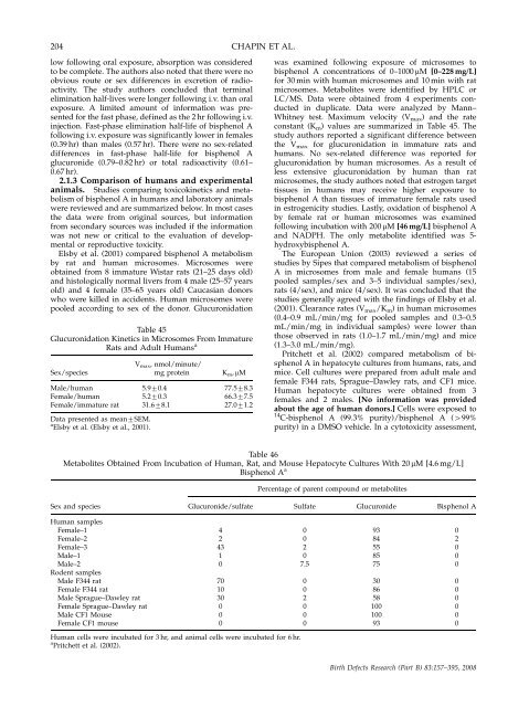
Table 45<br />
Glucur<strong>on</strong>idati<strong>on</strong> Kinetics in Microsomes From Immature<br />
Rats <strong>and</strong> Adult <strong>Human</strong>s a<br />
Sex/species<br />
Male/human<br />
Female/human<br />
Female/immature rat<br />
Data presented as mean7SEM.<br />
a Elsby et al. (Elsby et al., 2001).<br />
Vmax, nmol/minute/<br />
mg protein Km, mM<br />
5.970.4<br />
5.270.3<br />
31.678.1<br />
77.578.3<br />
66.377.5<br />
27.071.2<br />
was examined following exposure of microsomes to<br />
bisphenol A c<strong>on</strong>centrati<strong>on</strong>s of 0–1000 mM [0–228 mg/L]<br />
for 30 min with human microsomes <strong>and</strong> 10 min with rat<br />
microsomes. Metabolites were identified by HPLC or<br />
LC/MS. Data were obtained from 4 experiments c<strong>on</strong>ducted<br />
in duplicate. Data were analyzed by Mann–<br />
Whitney test. Maximum velocity (Vmax) <strong>and</strong> <strong>the</strong> rate<br />
c<strong>on</strong>stant (Km) values are summarized in Table 45. The<br />
study authors reported a significant difference between<br />
<strong>the</strong> V max for glucur<strong>on</strong>idati<strong>on</strong> in immature rats <strong>and</strong><br />
humans. No sex-related difference was reported for<br />
glucur<strong>on</strong>idati<strong>on</strong> by human microsomes. As a result of<br />
less extensive glucur<strong>on</strong>idati<strong>on</strong> by human than rat<br />
microsomes, <strong>the</strong> study authors noted that estrogen target<br />
tissues in humans may receive higher exposure to<br />
bisphenol A than tissues of immature female rats used<br />
in estrogenicity studies. Lastly, oxidati<strong>on</strong> of bisphenol A<br />
by female rat or human microsomes was examined<br />
following incubati<strong>on</strong> with 200 mM [46 mg/L] bisphenol A<br />
<strong>and</strong> NADPH. The <strong>on</strong>ly metabolite identified was 5hydroxybisphenol<br />
A.<br />
The European Uni<strong>on</strong> (2003) reviewed a series of<br />
studies by Sipes that compared metabolism of bisphenol<br />
A in microsomes from male <strong>and</strong> female humans (15<br />
pooled samples/sex <strong>and</strong> 3–5 individual samples/sex),<br />
rats (4/sex), <strong>and</strong> mice (4/sex). It was c<strong>on</strong>cluded that <strong>the</strong><br />
studies generally agreed with <strong>the</strong> findings of Elsby et al.<br />
(2001). Clearance rates (V max/Km) in human microsomes<br />
(0.4–0.9 mL/min/mg for pooled samples <strong>and</strong> 0.3–0.5<br />
mL/min/mg in individual samples) were lower than<br />
those observed in rats (1.0–1.7 mL/min/mg) <strong>and</strong> mice<br />
(1.3–3.0 mL/min/mg).<br />
Pritchett et al. (2002) compared metabolism of bisphenol<br />
A in hepatocyte cultures from humans, rats, <strong>and</strong><br />
mice. Cell cultures were prepared from adult male <strong>and</strong><br />
female F344 rats, Sprague–Dawley rats, <strong>and</strong> CF1 mice.<br />
<strong>Human</strong> hepatocyte cultures were obtained from 3<br />
females <strong>and</strong> 2 males. [No informati<strong>on</strong> was provided<br />
about <strong>the</strong> age of human d<strong>on</strong>ors.] Cells were exposed to<br />
14 C-bisphenol A (99.3% purity)/bisphenol A (499%<br />
purity) in a DMSO vehicle. In a cytotoxicity assessment,<br />
Table 46<br />
Metabolites Obtained From Incubati<strong>on</strong> of <strong>Human</strong>, Rat, <strong>and</strong> Mouse Hepatocyte Cultures With 20 mM [4.6 mg/L]<br />
Bisphenol A a<br />
Percentage of parent compound or metabolites<br />
Sex <strong>and</strong> species Glucur<strong>on</strong>ide/sulfate Sulfate Glucur<strong>on</strong>ide Bisphenol A<br />
<strong>Human</strong> samples<br />
Female–1 4 0 93 0<br />
Female–2 2 0 84 2<br />
Female–3 43 2 55 0<br />
Male–1 1 0 85 0<br />
Male–2 0 7.5 75 0<br />
Rodent samples<br />
Male F344 rat 70 0 30 0<br />
Female F344 rat 10 0 86 0<br />
Male Sprague–Dawley rat 30 2 58 0<br />
Female Sprague–Dawley rat 0 0 100 0<br />
Male CF1 Mouse 0 0 100 0<br />
Female CF1 mouse 0 0 93 0<br />
<strong>Human</strong> cells were incubated for 3 hr, <strong>and</strong> animal cells were incubated for 6 hr.<br />
a Pritchett et al. (2002).<br />
Birth Defects Research (Part B) 83:157–395, 2008


