

Monograph on the Potential Human Reproductive and ... - OEHHA
Monograph on the Potential Human Reproductive and ... - OEHHA
Monograph on the Potential Human Reproductive and ... - OEHHA

You also want an ePaper? Increase the reach of your titles
YUMPU automatically turns print PDFs into web optimized ePapers that Google loves.
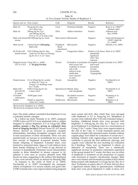
226 CHAPIN ET AL.<br />
Table 58<br />
In Vivo Genetic Toxicity Studies of Bisphenol A<br />
Species <strong>and</strong> sex Dose (route) Cells Endpoint Results Reference<br />
Male rat 85 mg/kg bw/day Germ Dominant lethality Negative B<strong>on</strong>d et al. (1980) a,b<br />
for 5 days (i.p.) (abstract <strong>on</strong>ly)<br />
Male rat 200 mg/kg bw (i.p.) DNA Adduct formati<strong>on</strong> Positive Atkins<strong>on</strong> <strong>and</strong><br />
<strong>and</strong> 200 mg/kg bw Roy (1995b)<br />
for 4, 8, 12, or 16 days (oral)<br />
Male <strong>and</strong> female 500–2000 mg/kg bw (oral) B<strong>on</strong>e marrow Micr<strong>on</strong>uclei Negative Gudi <strong>and</strong> Krsmanovic<br />
mouse (1999) a ; Shell Oil<br />
Co. (1999) b<br />
Male mouse 1 mmol/kg bw [228 mg/kg Peripheral Micr<strong>on</strong>uclei Negative Masuda et al. (2005)<br />
bw] (oral) blood<br />
reticulocyte<br />
20–22-day-old 0.02–0.100 mg/kg bw/day Oocyte C<strong>on</strong>gressi<strong>on</strong> failure Positive at all doses; Hunt et al. (2003)<br />
female mouse (oral) for 6–8 days or 0.02 mg/ statistically<br />
kg bw for 3, 5, or 7 days significant<br />
with 7-day<br />
exposure<br />
Pregnant mouse 0.4 mg/day s.c. pellet Oocyte Evaluati<strong>on</strong> of pachytene Incomplete synapsis, Susiarjo et al. (2007)<br />
GD 11.5–18.5 [B20 mg/kg bw/day] fetal oocyte <strong>and</strong> end-to-end<br />
of ploidy in oocytes associati<strong>on</strong><br />
<strong>and</strong> 2-cell of sister<br />
embryos from chromatids,<br />
adults that mhyperploidy<br />
were exposed<br />
in utero<br />
Female mouse 0.2 or 20 mg/kg bw acutely Oocyte Aneuploidy Negative Pacchierotti et al.<br />
or daily for 7 days or (2007)<br />
0.4 mg/L in drinking water<br />
for 7 weeks<br />
Male (102/ 0.002–0.2 mg/kg bw for Spermatocyte Meiotic delay Negative Pacchierotti et al.<br />
ElxC3H/El) 6 days (oral) <strong>and</strong> aneuploidy (2007)<br />
F 1 mouse<br />
Drosophila 10,000 ppm (oral) Offspring Sex-linked recessive Negative Foureman et al.<br />
melanogaster lethal test (1994) a,b<br />
Turbot 50 ppb in aquarium Erythrocyte Micr<strong>on</strong>uclei Positive Bolognesi et al.<br />
water for 2 weeks (2006)<br />
a Reviewed by Haight<strong>on</strong> et al. (2002).<br />
b Reviewed by European-Uni<strong>on</strong> (2003).<br />
days. The study authors c<strong>on</strong>cluded that bisphenol A was<br />
a potential meiotic aneugen.<br />
In a follow-up study (Susiarjo et al., 2007), pregnant<br />
C57Bl/6 mice <strong>on</strong> GD 11.5 were implanted with s.c. pellets<br />
designed to release bisphenol A 0 or 0.4 mg/day. [The<br />
authors assume a 20 g bw, giving an estimated dose<br />
level of 20 lg/kg bw/day.] Oocytes from GD 18.5 female<br />
fetuses showed an increase in pachytene synaptic<br />
abnormalities including incomplete synapsis <strong>and</strong> endto-end<br />
associati<strong>on</strong>s of sister chromatids. There was also<br />
paradoxically an increase in recombinant foci in pachytene<br />
oocytes of bisphenol A-exposed females. Some<br />
female offspring of bisphenol A-treated dams were<br />
fostered to untreated dams. Eggs or 2-cell embryos from<br />
<strong>the</strong>se female offspring at 4–5 weeks of age showed an<br />
increase in hyperploidy. Pachytene oocyte abnormalities<br />
similar to those identified in fetuses exposed to bisphenol<br />
A were seen in oocytes obtained from ERb<br />
knock-out mice, suggesting to <strong>the</strong> authors that bisphenol<br />
A may exert adverse effects <strong>on</strong> meiosis by blocking ERb.<br />
In resp<strong>on</strong>se to <strong>the</strong> study of Hunt et al. (2003),<br />
Pacchierotti et al. (2007) investigated <strong>the</strong> aneugenic<br />
effects of bisphenol A in mouse somatic <strong>and</strong> germ cells.<br />
C57Bl/6 female mice were superovulated using pregnant<br />
mare serum <strong>and</strong> hCG after which <strong>the</strong>y were gavaged<br />
with bisphenol A 0.2 or 20 mg/kg bw. Metaphase II<br />
oocytes were collected after 17 hr <strong>and</strong> evaluated using Cb<strong>and</strong>ing.<br />
Additi<strong>on</strong>al female mice were gavaged with<br />
bisphenol A 0.04 mg/kg bw/day for 7 days or were<br />
given bisphenol A in drinking water at a c<strong>on</strong>centrati<strong>on</strong> of<br />
0.4 mg/L for 7 weeks. These mice were superovulated at<br />
<strong>the</strong> end of <strong>the</strong> 7-day or 7-week treatment period <strong>and</strong><br />
housed overnight with untreated males. Females without<br />
vaginal plugs were killed for evaluati<strong>on</strong> of oocytes by Cb<strong>and</strong>ing.<br />
Females with vaginal plugs were treated with<br />
colchicine to prevent <strong>the</strong> first embry<strong>on</strong>ic cleavage, <strong>and</strong><br />
zygotes were collected <strong>the</strong> next morning for evaluati<strong>on</strong><br />
by C-b<strong>and</strong>ing. There were no bisphenol A effects <strong>on</strong><br />
inducti<strong>on</strong> of aneuploidy. There was a statistically<br />
significant increase in premature centromere separati<strong>on</strong><br />
in <strong>the</strong> group treated for 7 weeks, but <strong>the</strong>re was no effect<br />
of bisphenol A treatment <strong>on</strong> <strong>the</strong> proporti<strong>on</strong> of zygotes<br />
with structural or numeric chromosome changes. Male<br />
mice were treated with bromodeoxyuridine 8 days before<br />
being treated with bisphenol A 0.2 mg/kg bw/day for 6<br />
days. Evaluati<strong>on</strong> of sperm after 21–25 days did not show<br />
a significant mitotic delay in spermatocytes. Additi<strong>on</strong>al<br />
male mice were given bisphenol A orally at doses of 0,<br />
Birth Defects Research (Part B) 83:157–395, 2008


