

Monograph on the Potential Human Reproductive and ... - OEHHA
Monograph on the Potential Human Reproductive and ... - OEHHA
Monograph on the Potential Human Reproductive and ... - OEHHA

Create successful ePaper yourself
Turn your PDF publications into a flip-book with our unique Google optimized e-Paper software.
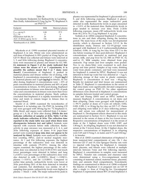
Table 26<br />
Toxicokinetic Endpoints for Radioactivity in Lactating<br />
Rats Orally Administered 0.5 mg/kg bw 14 C-Bisphenol A<br />
<strong>on</strong> PND 11 a<br />
Endpoint Milk Maternal plasma<br />
Cmax, mg-eq/L 4.46 27.2<br />
T max, hr 8 4<br />
Eliminati<strong>on</strong> half-life, hr 26 31<br />
AUC (0–48 hr), mg-eq � hr/L) 156 689<br />
a Kurebayashi et al. (2005).<br />
Miyakoda et al. (1999) examined placental transfer of<br />
bisphenol A in rats. Wistar rats were administered an<br />
oral dose of bisphenol A (99% purity) at 10 mg/kg bw <strong>on</strong><br />
GD 19. Blood was collected <strong>and</strong> fetuses were removed at<br />
1, 3, <strong>and</strong> 24 hr following dosing. Bisphenol A c<strong>on</strong>centrati<strong>on</strong>s<br />
were measured in plasma <strong>and</strong> fetuses by GC/MS.<br />
[A statement in Figure 3 of <strong>the</strong> study indicated that<br />
values were <strong>the</strong> means of 5 or 7 experiments; it is<br />
possible <strong>the</strong> authors meant that 5 or 7 dams were<br />
dosed.] C<strong>on</strong>centrati<strong>on</strong>s of bisphenol A peaked in<br />
maternal plasma <strong>and</strong> fetuses within 1 hr of dosing, with<br />
bisphenol A c<strong>on</strong>centrati<strong>on</strong>s measured at B34 ppb [lg/L]<br />
in maternal plasma <strong>and</strong> 11 ppb [lg/kg] in fetuses. At 3 hr<br />
after dosing, bisphenol A c<strong>on</strong>centrati<strong>on</strong>s were B10% of<br />
peak c<strong>on</strong>centrati<strong>on</strong>s in maternal plasma <strong>and</strong> 40% of peak<br />
c<strong>on</strong>centrati<strong>on</strong>s in fetuses. At 24 hr post-dosing, bisphenol<br />
A c<strong>on</strong>centrati<strong>on</strong>s in fetuses were detected at 70% of peak<br />
value <strong>and</strong> c<strong>on</strong>centrati<strong>on</strong>s in fetuses were more than twice<br />
<strong>the</strong> c<strong>on</strong>centrati<strong>on</strong>s in maternal plasma. Study authors<br />
c<strong>on</strong>cluded that bisphenol A is rapidly transferred to <strong>the</strong><br />
fetus <strong>and</strong> tends to remain l<strong>on</strong>ger in fetuses than in<br />
maternal blood.<br />
Snyder et al. (2000) examined <strong>the</strong> toxicokinetics of<br />
bisphenol A in lactating rats. On PND 14, lactating CD<br />
rats were gavaged with 100 mg/kg bw 14 C-bisphenol A.<br />
Milk, blood, <strong>and</strong> organs were collected from 2–4 dams/<br />
group at 1, 8, 24, or 26 hr after dosing. [While <strong>the</strong> text<br />
indicates collecti<strong>on</strong> of samples at 26 hr, Table 3 of <strong>the</strong><br />
study indicates collecti<strong>on</strong> at 24 hr. The collecti<strong>on</strong> time<br />
reported in <strong>the</strong> study table was used when <strong>the</strong>re were<br />
discrepancies between text <strong>and</strong> table.] Animals were<br />
injected with oxytocin before milk collecti<strong>on</strong>. Radioactivity<br />
in pup carcasses was measured at 2, 4, 6, <strong>and</strong><br />
24 hr following exposure of dams; 8–16 pups/time<br />
period were examined [pup data does not appear to be<br />
analyzed by litter]. Samples were analyzed by scintillati<strong>on</strong><br />
counting, HPLC, <strong>and</strong>/or nuclear magnetic res<strong>on</strong>ance.<br />
At 1 <strong>and</strong> 8 hr following exposure, <strong>the</strong> highest<br />
percentage of <strong>the</strong> radioactive dose was detected in<br />
intestine with c<strong>on</strong>tents (75–83%). Am<strong>on</strong>g <strong>the</strong> o<strong>the</strong>r<br />
organs examined, <strong>the</strong> highest percentage of <strong>the</strong> radioactive<br />
dose was detected in liver (0.38–0.74%) <strong>and</strong> much<br />
lower percentages were detected in kidney <strong>and</strong> lung<br />
(r0.02%). Low percentages of <strong>the</strong> radioactive dose were<br />
also detected in milk (r0.0020%), blood (B0.006%),<br />
plasma (B0.01%), <strong>and</strong> fat (r0.004%). Compared to<br />
earlier time periods, radioactivity levels were lower at<br />
24 hr post-dosing (26% of <strong>the</strong> dose detected in intestine<br />
<strong>and</strong> c<strong>on</strong>tents), but distributi<strong>on</strong> was similar. At all 3<br />
sampling time points, radioactivity levels were highest in<br />
plasma 4 blood 4 milk. The major radioactivity peak in<br />
Birth Defects Research (Part B) 83:157–395, 2008<br />
BISPHENOL A<br />
189<br />
plasma was represented by bisphenol A glucur<strong>on</strong>ide at 1,<br />
8, <strong>and</strong> 26 hr following exposure. Bisphenol A glucur<strong>on</strong>ide<br />
also represented <strong>the</strong> major radioactive peak<br />
detected in milk. Radioactivity levels in pups amounted<br />
to o0.01% of <strong>the</strong> maternal dose. Radioactivity levels in<br />
pups tended to increase over time. From 2–24 hr<br />
following exposure, mean7SD radioactivity levels rose<br />
from 44724 to 78711 mg bisphenol A eq/pup.<br />
Yoshida et al. (2004) compared bisphenol A c<strong>on</strong>centrati<strong>on</strong>s<br />
in rats <strong>and</strong> <strong>the</strong>ir offspring during <strong>the</strong> lactati<strong>on</strong><br />
period. The main focus of <strong>the</strong> study was developmental<br />
toxicity, which is discussed in Secti<strong>on</strong> 3.2.3.2. In <strong>the</strong><br />
distributi<strong>on</strong> study, D<strong>on</strong>ryu rats (12–19/group) were<br />
gavaged with bisphenol A at 0 (carboxymethylcellulose<br />
soluti<strong>on</strong>), 0.006, or 6 mg/kg bw/day from GD 2 to <strong>the</strong><br />
day before weaning (21 days post-delivery). Bisphenol A<br />
c<strong>on</strong>centrati<strong>on</strong>s were measured in maternal <strong>and</strong> pup<br />
serum, milk, <strong>and</strong> pup liver by GC/MS <strong>on</strong> PND 10, 14,<br />
<strong>and</strong>/or 21. Milk samples were obtained from pup<br />
stomachs. Pup serum <strong>and</strong> liver samples were pooled.<br />
Two to six dams/litter were examined in each dose<br />
group <strong>and</strong> time period. Samples of tap water, drinking<br />
water from plastic c<strong>on</strong>tainers, <strong>and</strong> feed were measured<br />
for bisphenol A c<strong>on</strong>tent by HPLC. Bisphenol A was not<br />
detected in fresh tap water but was detected at B3 mg/L<br />
following storage of that water in plastic c<strong>on</strong>tainers.<br />
Bisphenol A c<strong>on</strong>centrati<strong>on</strong> in feed was B40 mg/kg.<br />
Results for maternal <strong>and</strong> fetal tissues are summarized<br />
in Table 27. Bisphenol A c<strong>on</strong>centrati<strong>on</strong>s in <strong>the</strong> serum of<br />
high-dose-dams were significantly elevated compared to<br />
<strong>the</strong> c<strong>on</strong>trol group <strong>on</strong> PND 21. No o<strong>the</strong>r significant<br />
differences were observed in bisphenol A c<strong>on</strong>centrati<strong>on</strong>s<br />
in samples between treated <strong>and</strong> c<strong>on</strong>trol groups.<br />
Kim <strong>and</strong> Huang (2003) used an HPLC method to<br />
measure bisphenol A c<strong>on</strong>centrati<strong>on</strong>s in rat dams <strong>and</strong><br />
<strong>the</strong>ir offspring. Dams were gavaged with bisphenol A<br />
(499.7% purity) at doses of 0 (corn oil vehicle), 0.002,<br />
0.020, 0.200, 2, or 20 mg/kg bw/day <strong>on</strong> GD 7–17. Dams<br />
<strong>and</strong> offspring were killed at 21 days following parturiti<strong>on</strong>,<br />
<strong>and</strong> serum was collected for measurement of<br />
bisphenol A. Development effects observed in this study<br />
are summarized in Secti<strong>on</strong> 3.2.1.1. Bisphenol A was not<br />
detected in <strong>the</strong> serum of dams at <strong>the</strong> two lowest doses.<br />
Respective c<strong>on</strong>centrati<strong>on</strong>s of bisphenol A in <strong>the</strong> serum of<br />
dams at <strong>the</strong> 3 highest doses were 0.900, 0.987, <strong>and</strong><br />
1.00 mg/L. In offspring, bisphenol A was not detected in<br />
serum at <strong>the</strong> 3 lowest doses. At <strong>the</strong> 2 highest doses, <strong>the</strong><br />
respective c<strong>on</strong>centrati<strong>on</strong>s of bisphenol A in offspring<br />
were 0.69 <strong>and</strong> 0.74 mg/L in males <strong>and</strong> 0.71 <strong>and</strong> 0.82 mg/<br />
L in females.<br />
Shin et al. (2002) examined eliminati<strong>on</strong> of bisphenol A<br />
from maternal–fetal compartments of rats. On 1 day<br />
between GD 17 <strong>and</strong> 19, four Sprague–Dawley rats were<br />
i.v. injected with 2 mg/kg bw bisphenol A. Amniotic<br />
fluid, placenta, <strong>and</strong> fetuses were collected at multiple<br />
intervals between 5 min <strong>and</strong> 8 hr following injecti<strong>on</strong>.<br />
Bisphenol A c<strong>on</strong>centrati<strong>on</strong>s in samples were measured<br />
by HPLC. Transfer rate c<strong>on</strong>stants <strong>and</strong> clearance rates<br />
were determined using a five-compartment model<br />
c<strong>on</strong>sisting of maternal central, maternal tissue, placental,<br />
fetal, <strong>and</strong> amniotic fluid compartments. Toxicokinetic<br />
findings are summarized in Moors et al. (2006) evaluated<br />
<strong>the</strong> kinetics of bisphenol A in pregnant rats <strong>on</strong> GD 18<br />
after a single i.v. dose of 10 mg/kg bw. Unc<strong>on</strong>jugated<br />
bisphenol A represented almost 80% of total bisphenol A


