

Monograph on the Potential Human Reproductive and ... - OEHHA
Monograph on the Potential Human Reproductive and ... - OEHHA
Monograph on the Potential Human Reproductive and ... - OEHHA

You also want an ePaper? Increase the reach of your titles
YUMPU automatically turns print PDFs into web optimized ePapers that Google loves.
*<br />
individuals with <strong>the</strong> SULT1A1 2 enzyme (6.3178.91 mg/<br />
L). Adjustment for possible bisphenol A exposure<br />
through vinyl wrap use also did not result in significant<br />
differences between <strong>the</strong> 2 groups. The study authors<br />
c<strong>on</strong>cluded that additi<strong>on</strong>al enzymes involved in bisphenol<br />
A metabolism should be studied to determine<br />
possible sensitivity differences.<br />
One animal study dem<strong>on</strong>strated sex-related differences<br />
in sulfati<strong>on</strong>. Male versus female Sprague–Dawley<br />
<strong>and</strong> F344 rats were found to produce higher amounts of a<br />
bisphenol A glucur<strong>on</strong>ide/sulfate c<strong>on</strong>jugate (Pritchett<br />
et al., 2002).<br />
As noted in Table 7, <strong>on</strong>e human study reported B2fold<br />
higher blood bisphenol A c<strong>on</strong>centrati<strong>on</strong>s in Japanese<br />
men than women (Takeuchi <strong>and</strong> Tsutsumi, 2002). Based<br />
<strong>on</strong> positive correlati<strong>on</strong> between serum bisphenol A <strong>and</strong><br />
testoster<strong>on</strong>e c<strong>on</strong>centrati<strong>on</strong>s, authors speculated that sexrelated<br />
differences in bisphenol A c<strong>on</strong>centrati<strong>on</strong>s might<br />
be due to <strong>and</strong>rogen-related metabolism (Takeuchi <strong>and</strong><br />
Tsutsumi, 2002). There are no known human studies<br />
showing inter-individual or sex-related variati<strong>on</strong>s in<br />
metabolism that could lead to higher bisphenol A<br />
c<strong>on</strong>centrati<strong>on</strong>s in blood. Experimental animal studies<br />
have not c<strong>on</strong>sistently dem<strong>on</strong>strated higher c<strong>on</strong>centrati<strong>on</strong>s<br />
of bisphenol A or radioactive dose in <strong>on</strong>e sex<br />
(Pottenger et al., 2000; Kurebayashi et al., 2005). In Wistar<br />
rats orally administered 1 mg bisphenol A every 2 days<br />
Table 60<br />
<strong>Human</strong> Toxicokinetic Values for Total Bisphenol A Dose<br />
Endpoint Value Reference<br />
Oral absorpti<strong>on</strong>, % Z84% Völkel et al.(2002, 2005)<br />
Dermal absorpti<strong>on</strong>, in vitro, % B10% European-Uni<strong>on</strong> (2003)<br />
T max, min 80 Völkel et al. (2002)<br />
Eliminati<strong>on</strong> half-life, hr 4–5.4 Völkel et al. (2002, 2005)<br />
BISPHENOL A<br />
229<br />
for 2 or 4 weeks, liver microsomal UDPGT activity<br />
toward 17b-estradiol <strong>and</strong> testoster<strong>on</strong>e <strong>and</strong> expressi<strong>on</strong> of<br />
UGT2B1 protein <strong>and</strong> mRNA were reduced in males but<br />
not females (Shibata et al., 2002). One study reported an<br />
B3-fold higher c<strong>on</strong>centrati<strong>on</strong> of blood bisphenol A in<br />
male than in female Wistar–Imamichi rats that were<br />
apparently not treated, but <strong>the</strong>re were was no sex-related<br />
difference in percent glucur<strong>on</strong>idated bisphenol A in<br />
serum (Takeuchi et al., 2004b). However, in an in vitro<br />
study c<strong>on</strong>ducted with hepatic microsomes, glucur<strong>on</strong>idati<strong>on</strong><br />
of bisphenol A <strong>and</strong> expressi<strong>on</strong> of UGT2B1 mRNA<br />
were higher in microsomes from female than male rats.<br />
As described in more detail in Secti<strong>on</strong> 2.1.2.3, <strong>on</strong>e study<br />
showed reduced capacity to glucur<strong>on</strong>idate bisphenol A<br />
in livers from pregnant than in n<strong>on</strong>-pregnant rats (Inoue<br />
et al., 2004).<br />
2.6 Summary of General Toxicology <strong>and</strong> Biologic<br />
Effects<br />
Analytical c<strong>on</strong>siderati<strong>on</strong>s. Free c<strong>on</strong>centrati<strong>on</strong>s of<br />
BPA measured in various matrices can be affected by<br />
analytic techniques <strong>and</strong> methodology. Free bisphenol A<br />
c<strong>on</strong>taminati<strong>on</strong> from reagents <strong>and</strong> plastic ware may<br />
c<strong>on</strong>tribute to <strong>the</strong> measured free c<strong>on</strong>centrati<strong>on</strong> of bisphenol<br />
A (Tsukioka et al., 2004; Völkel et al., 2005).<br />
Analytical techniques employed may incorrectly overestimate<br />
<strong>the</strong> free c<strong>on</strong>centrati<strong>on</strong> of measured bisphenol A.<br />
HPLC with ultraviolet, fluorescence, or electrochemical<br />
detecti<strong>on</strong> is unable to make definitive identificati<strong>on</strong> of<br />
bisphenol A or bisphenol A glucur<strong>on</strong>ides, because<br />
similar retenti<strong>on</strong> times may occur for <strong>the</strong> metabolites of<br />
o<strong>the</strong>r endogenous <strong>and</strong> exogenous compounds (Völkel<br />
et al., 2005). Bisphenol A glucur<strong>on</strong>ide can also be<br />
hydrolyzed <strong>and</strong> in some cases degraded to unknown<br />
comp<strong>on</strong>ents ei<strong>the</strong>r in acidic or basic pH soluti<strong>on</strong>s of<br />
diluted urine, adding ano<strong>the</strong>r potential source of error in<br />
<strong>the</strong> measurement of sample levels of bisphenol A <strong>and</strong> its<br />
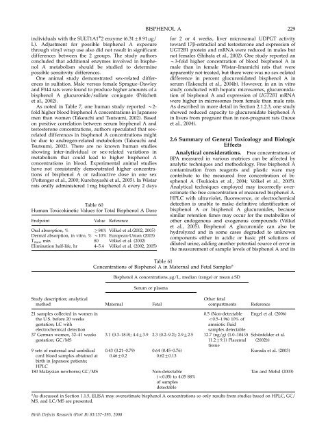
Table 61<br />
C<strong>on</strong>centrati<strong>on</strong>s of Bisphenol A in Maternal <strong>and</strong> Fetal Samples a<br />
Bisphenol A c<strong>on</strong>centrati<strong>on</strong>s, mg/L, median (range) or mean7SD<br />
Serum or plasma<br />
Study descripti<strong>on</strong>; analytical<br />
method Maternal Fetal<br />
O<strong>the</strong>r fetal<br />
compartments Reference<br />
21 samples collected in women in 0.5 (N<strong>on</strong>-detectable<br />
<strong>the</strong> U.S. before 20 weeks o0.5–1.96) 10% of<br />
gestati<strong>on</strong>; LC with amniotic fluid<br />
electrochemical detecti<strong>on</strong> samples detectable<br />
37 German women, 32–41 weeks 3.1 (0.3–18.9); 4.473.9 2.3 (0.2–9.2); 2.972.5 12.7 (ng/g) (1.0–104.9)<br />
gestati<strong>on</strong>; GC/MS 11.279.1) Placental<br />
tissue<br />
9 sets of maternal <strong>and</strong> umbilical 0.43 (0.21–0.79) 0.64 (0.45–0.76)<br />
cord blood samples obtained at 0.4670.2 0.6270.13<br />
birth in Japanese patients;<br />
HPLC<br />
180 Malaysian newborns; GC/MS N<strong>on</strong>-detectable<br />
(o0.05) to 4.05 88%<br />
of samples<br />
detectable<br />
Engel et al. (2006)<br />
Schönfelder et al.<br />
(2002b)<br />
Kuroda et al. (2003)<br />
Tan <strong>and</strong> Mohd (2003)<br />
a As discussed in Secti<strong>on</strong> 1.1.5, ELISA may overestimate bisphenol A c<strong>on</strong>centrati<strong>on</strong>s so <strong>on</strong>ly results from studies based <strong>on</strong> HPLC, GC/<br />
MS, <strong>and</strong> LC/MS are presented.<br />
Birth Defects Research (Part B) 83:157–395, 2008


