

Monograph on the Potential Human Reproductive and ... - OEHHA
Monograph on the Potential Human Reproductive and ... - OEHHA
Monograph on the Potential Human Reproductive and ... - OEHHA

Create successful ePaper yourself
Turn your PDF publications into a flip-book with our unique Google optimized e-Paper software.
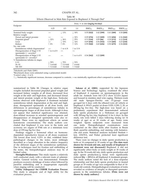
342 CHAPIN ET AL.<br />
Endpoint<br />
Table 88<br />
Effects Observed in Male Rats Exposed to Bisphenol A Through Diet a<br />
Dose, % in diet [mg/kg bw/day]<br />
0.25 0.5 1.0 BMD10 BMDL10 BMD1SD BMDL1SD<br />
Terminal body weight<br />
Relative weight<br />
2 k 13% k 18% 0.55 [522] 0.42 [399] 0.41 [389] 0.30 [285]<br />
Dorsal <strong>and</strong> lateral prostate 2 2 k 32% 0.29 [276] 0.22 [209] 0.52 [494] 0.36 [342]<br />
Preputial gl<strong>and</strong> b<br />
k 22% k 26% k 25% 0.13 [124] 0.09 [86] 0.18 [171] 0.12 [114]<br />
Liver 2 k 10% k 14% 0.69 [656] 0.56 [532] 0.30 [285] 0.23 [218]<br />
Kidney<br />
No. rats with:<br />
m 8% m 8% m 12% 0.99 [940] 0.69 [656] 0.50 [475] 0.34 [323]<br />
Seminiferous tubule degenerati<strong>on</strong> c 2 m to 6/8 m to 5/8<br />
Disorganizati<strong>on</strong> of Stage I–VI<br />
spermatids (1 severity) c<br />
m to 4 of 8 2 2<br />
Disorganizati<strong>on</strong> of Stage I–VI 2 2 m to 6 of 8 0.36 [342] 0.22 [209]<br />
spermatids (21 severity) c<br />
% Seminiferous tubules in stages<br />
I–VI k 59% k 70% k 53%<br />
IX–XI m 3.4-fold m 5.2-fold m 4-fold<br />
XII–XIV m 3.2-fold m 3.6-fold m 3-fold<br />
a<br />
Takahashi <strong>and</strong> Oishi (2001).<br />
b<br />
Benchmark doses were estimated using a polynomial model.<br />
c<br />
C<strong>on</strong>trol value 5 0of8. m,k Statistically significant increase, decrease compared to c<strong>on</strong>trols; 2no statistically significant effect compared to c<strong>on</strong>trols.<br />
summarized in Table 88. Changes in relative organ<br />
weights included decreased preputial gl<strong>and</strong> weight <strong>and</strong><br />
increased kidney weights at all doses, decreased liver<br />
weight at <strong>the</strong> mid- <strong>and</strong> high-dose, <strong>and</strong> decreased dorsal<br />
<strong>and</strong> lateral prostate weight at <strong>the</strong> high-dose. Testicular<br />
lesi<strong>on</strong>s observed with bisphenol A treatment included<br />
seminiferous tubule degenerati<strong>on</strong> at <strong>the</strong> mid <strong>and</strong> highdose,<br />
disorganized spermatids at all dose levels, <strong>and</strong><br />
differences in percentages of seminiferous tubules in<br />
spermatogenic stages at all dose levels. Although it does<br />
not appear that statistical significance was attained,<br />
dose-related increases in arrested spermatogenesis <strong>and</strong><br />
disappearance of el<strong>on</strong>gated spermatids were also reported.<br />
There were no significant effects <strong>on</strong> serum<br />
testoster<strong>on</strong>e c<strong>on</strong>centrati<strong>on</strong>s. The study authors c<strong>on</strong>cluded<br />
that bisphenol A was toxic to <strong>the</strong> testis <strong>and</strong><br />
accessory sex organs of F344 rats at a minimum toxic<br />
dose of 235 mg/kg bw/day.<br />
Findings suggest a horm<strong>on</strong>al effect <strong>on</strong> horm<strong>on</strong>edependent<br />
reproductive tissues at all doses examined.<br />
The lowest dose level, 0.25% in diet, exhibited histopathological<br />
changes in <strong>the</strong> testes, most strikingly<br />
described as a large alterati<strong>on</strong> in <strong>the</strong> relative frequency<br />
of <strong>the</strong> different stages of <strong>the</strong> seminiferous epi<strong>the</strong>lium.<br />
Due to techniques used for fixati<strong>on</strong> <strong>and</strong> embedding of<br />
<strong>the</strong> testes, <strong>the</strong> histopathological analyses may be of<br />
limited value.<br />
Strengths/Weaknesses: This study reports a relatively<br />
well c<strong>on</strong>ducted study with a relevant route of administrati<strong>on</strong>.<br />
General toxicity was dem<strong>on</strong>strated. Formalin<br />
produces excessive shrinkage of testes when followed by<br />
paraffin embedding <strong>and</strong> is inappropriate especially<br />
when staging will be c<strong>on</strong>ducted.<br />
Utility (Adequacy) for CERHR Evaluati<strong>on</strong> Process:<br />
This study is adequate <strong>and</strong> of high utility for <strong>the</strong><br />
evaluati<strong>on</strong> process.<br />
Sakaue et al. (2001), supported by <strong>the</strong> Japanese<br />
Science <strong>and</strong> Technology Agency, examined <strong>the</strong> effect<br />
of bisphenol A exposure <strong>on</strong> spermatogenesis in <strong>the</strong><br />
adult rat. Animals were fed CE-2 chow (CLEA Japan)<br />
<strong>and</strong> housed in stainless steel wire caging. Thirteen-week<br />
old male Sprague–Dawley rats (5/group) were<br />
gavaged for 6 days with <strong>the</strong> ethanol/corn oil vehicle or<br />
bisphenol A (99.6% purity) at doses 0.020, 0.200, 2, 20, or<br />
200 mg/kg bw/day. The high-dose was based <strong>on</strong> a<br />
preliminary experiment that dem<strong>on</strong>strated reduced<br />
daily sperm producti<strong>on</strong> in a Holtzman rat gavaged<br />
with 200 mg/kg bw/day bisphenol A for 6 days. In this<br />
study, rats were killed 2 days following dosing (at 14<br />
weeks of age) or at 18 weeks of age. Testes were<br />
weighed. Sperm endpoints were measured from<br />
<strong>on</strong>e testis. Histopathological examinati<strong>on</strong>s were<br />
c<strong>on</strong>ducted <strong>on</strong> <strong>the</strong> o<strong>the</strong>r testis after fixati<strong>on</strong> in Bouin<br />
fluid, paraffin embedding, <strong>and</strong> staining with hematoxylin<br />
<strong>and</strong> eosin. Statistical analyses included Student ttest,<br />
ANOVA, <strong>and</strong> Fisher protected least significant<br />
difference test.<br />
There were no changes in daily sperm producti<strong>on</strong>/g<br />
testis at 14 compared to 18 weeks of age. [No data were<br />
shown for 14-week-old rats, <strong>and</strong> results of bisphenol A<br />
treatment were not discussed.] Bisphenol A did not<br />
significantly affect body or testis weight at 18 weeks of<br />
age. In <strong>the</strong> 18-week-old rats, daily sperm producti<strong>on</strong> <strong>and</strong><br />
daily sperm producti<strong>on</strong>/g tissue were significantly<br />
reduced [by B25%] in all bisphenol A treatment groups.<br />
The study authors noted <strong>the</strong> lack of a dose–resp<strong>on</strong>se<br />
relati<strong>on</strong>ship <strong>and</strong> that daily sperm producti<strong>on</strong> in treated<br />
groups at 18 weeks of age was comparable to that of 14week-old<br />
c<strong>on</strong>trols. Histopathological evaluati<strong>on</strong>s of testis<br />
revealed no evidence of atrophy or disrupted spermatogenesis<br />
in <strong>the</strong> seminiferous tubules. [Data were not<br />
shown by study authors.]<br />
Birth Defects Research (Part B) 83:157–395, 2008


