

Monograph on the Potential Human Reproductive and ... - OEHHA
Monograph on the Potential Human Reproductive and ... - OEHHA
Monograph on the Potential Human Reproductive and ... - OEHHA

Create successful ePaper yourself
Turn your PDF publications into a flip-book with our unique Google optimized e-Paper software.
Endpoint<br />
BISPHENOL A<br />
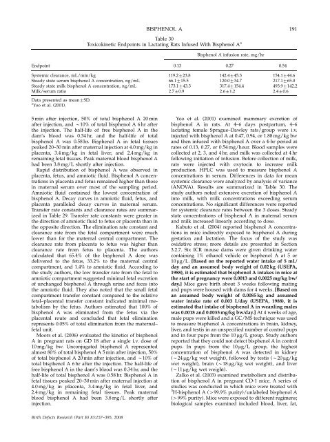
Table 30<br />
Toxicokinetic Endpoints in Lactating Rats Infused With Bisphenol A a<br />
0.13<br />
Bisphenol<br />
A infusi<strong>on</strong> rate, mg/hr<br />
Systemic clearance, mL/min/kg 119.2723.8 142.4745.3 154.1744.6<br />
Steady state serum bisphenol A c<strong>on</strong>centrati<strong>on</strong>, ng/mL 66.1715.5 120.0734.7 217.1765.0<br />
Steady state milk bisphenol A c<strong>on</strong>centrati<strong>on</strong>, ng/mL 173.1743.3 317.47154.4 493.97142.2<br />
Milk/serum ratio 2.770.9 2.671.2 2.470.6<br />
Data presented as mean7SD.<br />
a Yoo et al. (2001).<br />
5 min after injecti<strong>on</strong>, 50% of total bisphenol A 20 min<br />
after injecti<strong>on</strong>, <strong>and</strong> B10% of total bisphenol A 6 hr after<br />
<strong>the</strong> injecti<strong>on</strong>. The half-life of free bisphenol A in <strong>the</strong><br />
dam’s blood was 0.34 hr, <strong>and</strong> <strong>the</strong> half-life of total<br />
bisphenol A was 0.58 hr. Bisphenol A in fetal tissues<br />
peaked 20–30 min after maternal injecti<strong>on</strong> at 4.0 mg/kg in<br />
placenta, 3.4 mg/kg in fetal liver, <strong>and</strong> 2.4 mg/kg in<br />
remaining fetal tissues. Peak maternal blood bisphenol A<br />
had been 3.8 mg/L shortly after injecti<strong>on</strong>.<br />
Rapid distributi<strong>on</strong> of bisphenol A was observed in<br />
placenta, fetus, <strong>and</strong> amniotic fluid. Bisphenol A c<strong>on</strong>centrati<strong>on</strong>s<br />
in placenta <strong>and</strong> fetus remained higher than those<br />
in maternal serum over most of <strong>the</strong> sampling period.<br />
Amniotic fluid c<strong>on</strong>tained <strong>the</strong> lowest c<strong>on</strong>centrati<strong>on</strong> of<br />
bisphenol A. Decay curves in amniotic fluid, fetus, <strong>and</strong><br />
placenta paralleled decay curves in maternal serum.<br />
Transfer rate c<strong>on</strong>stants <strong>and</strong> clearance rates are summarized<br />
in Table 29. Transfer rate c<strong>on</strong>stants were greater in<br />
<strong>the</strong> directi<strong>on</strong> of amniotic fluid to fetus or placenta than in<br />
<strong>the</strong> opposite directi<strong>on</strong>. The eliminati<strong>on</strong> rate c<strong>on</strong>stant <strong>and</strong><br />
clearance rate from <strong>the</strong> fetal compartment were much<br />
lower than for <strong>the</strong> maternal central compartment. The<br />
clearance rate from placenta to fetus was higher than<br />
clearance rate from fetus to placenta. The authors<br />
calculated that 65.4% of <strong>the</strong> bisphenol A dose was<br />
delivered to <strong>the</strong> fetus, 33.2% to <strong>the</strong> maternal central<br />
compartment, <strong>and</strong> 1.4% to amniotic fluid. According to<br />
<strong>the</strong> study authors, <strong>the</strong> low transfer rate from <strong>the</strong> fetal to<br />
amniotic compartment suggested minimal fetal excreti<strong>on</strong><br />
of unchanged bisphenol A through urine <strong>and</strong> feces into<br />
<strong>the</strong> amniotic fluid. They also noted that <strong>the</strong> small fetal<br />
compartment transfer c<strong>on</strong>stant compared to <strong>the</strong> relative<br />
fetal–placental transfer c<strong>on</strong>stant indicated minimal metabolism<br />
by <strong>the</strong> fetus. Authors estimated that 100% of<br />
bisphenol A was eliminated from <strong>the</strong> fetus via <strong>the</strong><br />
placental route <strong>and</strong> c<strong>on</strong>cluded that fetal eliminati<strong>on</strong><br />
represents 0.05% of total eliminati<strong>on</strong> from <strong>the</strong> maternal–<br />
fetal unit.<br />
Moors et al. (2006) evaluated <strong>the</strong> kinetics of bisphenol<br />
A in pregnant rats <strong>on</strong> GD 18 after a single i.v. dose of<br />
10 mg/kg bw. Unc<strong>on</strong>jugated bisphenol A represented<br />
almost 80% of total bisphenol A 5 min after injecti<strong>on</strong>, 50%<br />
of total bisphenol A 20 min after injecti<strong>on</strong>, <strong>and</strong> B10% of<br />
total bisphenol A 6 hr after <strong>the</strong> injecti<strong>on</strong>. The half-life of<br />
free bisphenol A in <strong>the</strong> dam’s blood was 0.34 hr, <strong>and</strong> <strong>the</strong><br />
half-life of total bisphenol A was 0.58 hr. Bisphenol A in<br />
fetal tissues peaked 20–30 min after maternal injecti<strong>on</strong> at<br />
4.0 mg/kg in placenta, 3.4 mg/kg in fetal liver, <strong>and</strong><br />
2.4 mg/kg in remaining fetal tissues. Peak maternal<br />
blood bisphenol A had been 3.8 mg/L shortly after<br />
injecti<strong>on</strong>.<br />
Birth Defects Research (Part B) 83:157–395, 2008<br />
0.27<br />
0.54<br />
191<br />
Yoo et al. (2001) examined mammary excreti<strong>on</strong> of<br />
bisphenol A in rats. At 4–6 days postpartum, 4–6<br />
lactating female Sprague–Dawley rats/group were i.v.<br />
injected with bisphenol A at 0.47, 0.94, or 1.88 mg/kg bw<br />
<strong>and</strong> <strong>the</strong>n infused with bisphenol A over a 4-hr period at<br />
rates of 0.13, 0.27, or 0.54 mg/hour. Blood samples were<br />
collected at 2, 3, <strong>and</strong> 4 hr, <strong>and</strong> milk was collected at 4 hr<br />
following initiati<strong>on</strong> of infusi<strong>on</strong>. Before collecti<strong>on</strong> of milk,<br />
rats were injected with oxytocin to increase milk<br />
producti<strong>on</strong>. HPLC was used to measure bisphenol A<br />
c<strong>on</strong>centrati<strong>on</strong>s in serum. Differences in data for mean<br />
systemic clearance were analyzed by analysis of variance<br />
(ANOVA). Results are summarized in Table 30. The<br />
study authors noted extensive excreti<strong>on</strong> of bisphenol A<br />
into milk, with milk c<strong>on</strong>centrati<strong>on</strong>s exceeding serum<br />
c<strong>on</strong>centrati<strong>on</strong>s. No significant differences were reported<br />
for systemic clearance rates between <strong>the</strong> 3 doses. Steady<br />
state c<strong>on</strong>centrati<strong>on</strong>s of bisphenol A in maternal serum<br />
<strong>and</strong> milk increased linearly according to dose.<br />
Kabuto et al. (2004) reported bisphenol A c<strong>on</strong>centrati<strong>on</strong>s<br />
in mice indirectly exposed to bisphenol A during<br />
gestati<strong>on</strong> <strong>and</strong> lactati<strong>on</strong>. The focus of <strong>the</strong> study was<br />
oxidative stress; more details are presented in Secti<strong>on</strong><br />
3.2.7. Six ICR mouse dams were given drinking water<br />
c<strong>on</strong>taining 1% ethanol vehicle or bisphenol A at 5 or<br />
10 mg/L. [Based <strong>on</strong> <strong>the</strong> reported water intake of 5 mL/<br />
day <strong>and</strong> an assumed body weight of 0.02 kg (USEPA,<br />
1988), it is estimated that bisphenol A intakes in mice at<br />
<strong>the</strong> start of pregnancy were 0.0013 <strong>and</strong> 0.0025 mg/kg bw/<br />
day.] Mice gave birth about 3 weeks following mating<br />
<strong>and</strong> pups were housed with dams for 4 weeks. [Based <strong>on</strong><br />
an assumed body weight of 0.0085 kg <strong>and</strong> assumed<br />
water intake rate of 0.003 L/day (USEPA, 1988), it is<br />
estimated that intake of bisphenol A in weanling males<br />
was 0.0018 <strong>and</strong> 0.0035 mg/kg bw/day.] At 4 weeks of age,<br />
male pups were killed <strong>and</strong> a GC/MS technique was used<br />
to measure bisphenol A c<strong>on</strong>centrati<strong>on</strong>s in brain, kidney,<br />
liver, <strong>and</strong> testis in an unspecified number of c<strong>on</strong>trol pups<br />
<strong>and</strong> in four pups from <strong>the</strong> 10 mg/L group. Study authors<br />
reported that <strong>the</strong>y could not detect bisphenol A in c<strong>on</strong>trol<br />
pups. In pups from <strong>the</strong> 10 mg/L group, <strong>the</strong> highest<br />
c<strong>on</strong>centrati<strong>on</strong> of bisphenol A was detected in kidney<br />
(B24 mg/kg wet weight), followed by testis (B20 mg/kg<br />
wet weight), brain (B18 mg/kg wet weight), <strong>and</strong> liver<br />
(B11 mg/kg wet weight).<br />
Zalko et al. (2003) examined metabolism <strong>and</strong> distributi<strong>on</strong><br />
of bisphenol A in pregnant CD-1 mice. A series of<br />
studies was c<strong>on</strong>ducted in which mice were treated with<br />
3 H-bisphenol A (499.9% purity)/unlabeled bisphenol A<br />
(499% purity). Mice were exposed to different regimens;<br />
biological samples examined included blood, liver, fat,


