

Monograph on the Potential Human Reproductive and ... - OEHHA
Monograph on the Potential Human Reproductive and ... - OEHHA
Monograph on the Potential Human Reproductive and ... - OEHHA

You also want an ePaper? Increase the reach of your titles
YUMPU automatically turns print PDFs into web optimized ePapers that Google loves.
292 CHAPIN ET AL.<br />
has been informed that <strong>the</strong>re were 20–30/group (A.<br />
Soto, pers<strong>on</strong>al communicati<strong>on</strong>, March 2, 2007).] Fetal<br />
mammary gl<strong>and</strong>s were mounted whole or secti<strong>on</strong>ed to<br />
examine mammary gl<strong>and</strong> development in 36–40 offspring/group.<br />
Immunohistochemistry techniques were<br />
used to measure expressi<strong>on</strong> of Ki67 <strong>and</strong> Bax in mammary<br />
structures from 4–8 offspring/group. Mammary collagen<br />
localizati<strong>on</strong> was assessed using Mass<strong>on</strong> Trichrome stain<br />
in 6–17 mice/group. Expressi<strong>on</strong> of mRNA for ERa, ERb,<br />
adipocyte lipid binding protein, Col-l, <strong>and</strong> PPARg were<br />
measured by RT-PCR in mammary gl<strong>and</strong>s from 4–6<br />
offspring/group. Litter was accounted for in design <strong>and</strong><br />
analyses by assigning 1 individual/litter to each group or<br />
endpoint. Statistical analyses included t-tests, ANOVA,<br />
Mann–Whitney U n<strong>on</strong>-parametric tests, <strong>and</strong> w 2 tests.<br />
Morphometric analysis revealed significantly higher<br />
ductal area <strong>and</strong> extensi<strong>on</strong> in <strong>the</strong> bisphenol A group than<br />
in c<strong>on</strong>trols. In <strong>the</strong> c<strong>on</strong>trol group, females positi<strong>on</strong>ed next to<br />
two females in utero had significantly fewer branching<br />
points than females positi<strong>on</strong>ed next to 1 or 2 males; this<br />
difference was not observed in <strong>the</strong> bisphenol A group. In<br />
fetuses that were not positi<strong>on</strong>ed next to a male, significantly<br />
more branching points were observed in <strong>the</strong><br />
bisphenol A than in <strong>the</strong> c<strong>on</strong>trol group. C<strong>on</strong>trol females<br />
positi<strong>on</strong>ed next to 2 males had significantly larger<br />
epi<strong>the</strong>lial duct area than c<strong>on</strong>trol females not positi<strong>on</strong>ed<br />
next to a male; this difference was not observed in <strong>the</strong><br />
bisphenol A group. In bisphenol A-treated females positi<strong>on</strong>ed<br />
next to 1 male, ductal extensi<strong>on</strong> was significantly<br />
greater than in c<strong>on</strong>trol females positi<strong>on</strong>ed next to 1 male.<br />
In <strong>the</strong> bisphenol A group, epi<strong>the</strong>lial cells were less<br />
rounded, more evenly spaced, <strong>and</strong> more dense than<br />
in c<strong>on</strong>trols. Bisphenol A did not significantly affect Ki67<br />
(a proliferati<strong>on</strong> marker) expressi<strong>on</strong> in mammary epi<strong>the</strong>lium.<br />
Lumen formati<strong>on</strong> was observed in 6 of 16 c<strong>on</strong>trol<br />
mice <strong>and</strong> 0 of 10 bisphenol A-exposed mice. Significantly<br />
decreased numbers of Bax-positive (apoptotic) cells were<br />
observed in <strong>the</strong> inner epi<strong>the</strong>lial cord (not in c<strong>on</strong>tact with<br />
basement membrane) of bisphenol A-exposed than<br />
c<strong>on</strong>trol mice. Optical density of histological staining<br />
was significantly lower in <strong>the</strong> fat pad of <strong>the</strong> bisphenol Aexposed<br />
than c<strong>on</strong>trol group. Fat pads of <strong>the</strong> bisphenol A<br />
group compared to c<strong>on</strong>trol group were found to be<br />
significantly less cellular, c<strong>on</strong>tain more Bax-positive cells,<br />
<strong>and</strong> have more vacuoles at a distance o1 mm from <strong>the</strong><br />
epi<strong>the</strong>lial compartment. Study authors interpreted <strong>the</strong><br />
effect as increased epi<strong>the</strong>lial penetrati<strong>on</strong> <strong>and</strong> advanced<br />
maturati<strong>on</strong> of fat pads. No significant differences were<br />
observed for PPARg or adipocyte lipid binding protein<br />
mRNA expressi<strong>on</strong>. Density of collagen deposits was<br />
lower in <strong>the</strong> entire mammary gl<strong>and</strong> but higher in <strong>the</strong><br />
periductal stroma (within 10 mM of <strong>the</strong> epi<strong>the</strong>lium) of <strong>the</strong><br />
bisphenol A than <strong>the</strong> c<strong>on</strong>trol group. Bisphenol A<br />
exposure did not affect collagen type I, ERa, or ERb<br />
mRNA expressi<strong>on</strong>. ERa protein expressi<strong>on</strong> in <strong>the</strong> stroma<br />
was also unaffected by bisphenol A exposure. Study<br />
authors c<strong>on</strong>cluded that advanced maturati<strong>on</strong> of fat pad<br />
<strong>and</strong> changes in extracellular matrix may be <strong>the</strong> cause of<br />
altered growth, cell size, <strong>and</strong> lumen formati<strong>on</strong> in<br />
mammary epi<strong>the</strong>lium of mouse fetuses exposed to<br />
bisphenol A.<br />
Strengths/Weaknesses: Strengths of this study are <strong>the</strong><br />
rigor with which <strong>the</strong> measurements were made, <strong>and</strong> <strong>the</strong><br />
fact that <strong>the</strong> authors were trying to quantify endpoints<br />
that are difficult to measure (e.g., <strong>the</strong> relati<strong>on</strong>ship of <strong>the</strong><br />
stroma to <strong>the</strong> epi<strong>the</strong>lium). The relevance of <strong>the</strong> endpoints<br />
is a strength as is <strong>the</strong> low dose used. The single dose <strong>and</strong><br />
subcutaneous route of administrati<strong>on</strong> are weaknesses. A<br />
critical weakness is inappropriate statistical analysis of a<br />
complex study design that may have produced too many<br />
positive findings <strong>and</strong> a lack of statistical accounting for<br />
litter effects (i.e.,. 36–40 pups presented in Table 1 of<br />
study <strong>and</strong> <strong>on</strong>ly 20–30 litters treated).<br />
Utility (Adequacy) for CERHR Evaluati<strong>on</strong> Process:<br />
This study is inadequate for <strong>the</strong> evaluati<strong>on</strong> process<br />
because of insufficient c<strong>on</strong>trol for litter effects.<br />
H<strong>on</strong>ma et al. (2002), supported by <strong>the</strong> Japanese<br />
Ministry of Educati<strong>on</strong>, Culture, Sports, Sciences, <strong>and</strong><br />
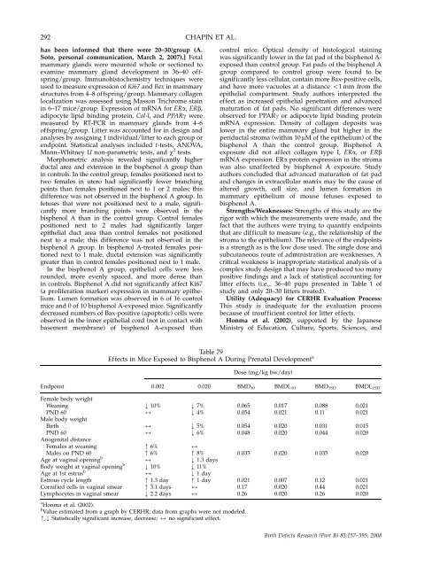
Table 79<br />
Effects in Mice Exposed to Bisphenol A During Prenatal Development a<br />
Dose (mg/kg bw/day)<br />
Endpoint 0.002 0.020 BMD10 BMDL10 BMD1SD BMDL1SD<br />
Female body weight<br />
Weaning k 10% k 7% 0.065 0.017 0.088 0.021<br />
PND 60<br />
Male body weight<br />
2 k 4% 0.054 0.021 0.11 0.021<br />
Birth 2 k 5% 0.054 0.020 0.031 0.015<br />
PND 60<br />
Anogenital distance<br />
2 k 6% 0.048 0.020 0.044 0.020<br />
Females at weaning m 6% 2<br />
Males <strong>on</strong> PND 60 m 6% m 8% 0.035 0.020 0.035 0.020<br />
Age at vaginal opening b<br />
2 k 1.3 days<br />
Body weight at vaginal opening b<br />
k 10% k 11%<br />
Age at 1st estrus b<br />
2 k 1 day<br />
Estrous cycle length m 1.3 day m 1 day 0.021 0.007 0.12 0.021<br />
Cornified cells in vaginal smear m 3.1 days 2 0.17 0.020 0.44 0.021<br />
Lymphocytes in vaginal smear k 2.2 days 2 0.26 0.020 0.26 0.020<br />
a H<strong>on</strong>ma et al. (2002).<br />
b Value estimated from a graph by CERHR; data from graphs were not modeled.<br />
m,k Statistically significant increase, decrease; 2 no significant effect.<br />
Birth Defects Research (Part B) 83:157–395, 2008


