Indice - Giornale Italiano di Medicina del Lavoro ed Ergonomia ...
Indice - Giornale Italiano di Medicina del Lavoro ed Ergonomia ...
Indice - Giornale Italiano di Medicina del Lavoro ed Ergonomia ...

You also want an ePaper? Increase the reach of your titles
YUMPU automatically turns print PDFs into web optimized ePapers that Google loves.
16 G Ital M<strong>ed</strong> Lav Erg 2006; 28:3, Suppl<br />
www.gimle.fsm.it<br />
5) Kühl WM, Bergsagel PL. Multiple myeloma: evolving genetic<br />
events and host interactions. Nat Rev Cancer 2002; 2: 175-187.<br />
6) Tafani A, Pregnolato A, Negri E et al. Diet and risk of lymphoid neoplasma<br />
and soft tissue sarcomas. Nutr Cancer 1997; 27: 256-260.<br />
7) Gramenzi A, Buttino I, D’Avanzo B, et al. Me<strong>di</strong>cal history and the<br />
risk of multiple myeloma. Br J Cancer 1991; 63: 769-772.<br />
8) Ende M. Multiple myeloma: a cluster in Virginia? Va M<strong>ed</strong> 1979;<br />
106: 115-116.<br />
9) Sonoda T, Ispida T, Mori M, et al. A case-control study of multiple<br />
myeloma in Japan: association with occupational factors. Asian Pac<br />
J Cancer Prev 2005; 6: 33-36.<br />
10) Demers PA, Vaughan TL, Köpsell et al. A case-control study of multiple<br />
myeloma and occupation. Am J Ind M<strong>ed</strong> 1993; 23: 629-639.<br />
11) Mester B, Nieters A, Deeg E et al. Occupation and malignant<br />
lymphoma: a population bas<strong>ed</strong> case control study in Germany. Occup<br />
Environ M<strong>ed</strong> 2006; 36: 17-26.<br />
12) Altieri A, Chen B, Bermejo JL et al. Familial risks and temporal incidence<br />
trends of multiple myeloma. Eur J Cancer 2006:in strampa<br />
13) Kyle RA, Finkelstein S, Elveback LR, et al. Incidente of monoclonal<br />
proteins in a Minnesota community with a cluster of multiple myeloma.<br />
Blood 1972; 40: 719-724.<br />
14) Kyle RA, Heath Jr CW, Carbone P. Multiple myeloma in spouses.<br />
Arch Intern M<strong>ed</strong> 1971; 127: 944-946.<br />
15) Kyle RA, Greipp PR. Multiple myeloma. Houses and spouses. Cancer<br />
1983; 51: 735-739.<br />
16) Linet MS. Chronic lymphocytic leukaemia and multiple myeloma in<br />
husband and wife. Am J M<strong>ed</strong> Sci 1984; 288: 21-24.<br />
17) Khuder SA, Mutgi AB. Meta-analyses of multiple myeloma and farming.<br />
Am J Ind M<strong>ed</strong> 1997; 32: 510-516.<br />
P-05<br />
32 P-POSTLABELING E POLIACRILAMIDE GEL ELETTROFORESI:<br />
CONFRONTO E APPLICAZIONE DELLE DUE TECNICHE<br />
PER IL RILEVAMENTO DEGLI ADDOTTI AL DNA<br />
B. Pappalar<strong>di</strong>1 , M.V. Policastro1 , P. Chiumarulo1 , G. Assennato1 ,<br />
P. Corsi2 1 Dip. <strong>di</strong> Me<strong>di</strong>cina Interna e Pubblica, Sez. <strong>di</strong> Me<strong>di</strong>cina <strong>del</strong> <strong>Lavoro</strong><br />
B. Ramazzini<br />
2 Dip. <strong>di</strong> Farmacologia e Fisiologia Umana<br />
Facoltà <strong>di</strong> Me<strong>di</strong>cina e Chirurgia, Università degli Stu<strong>di</strong> <strong>di</strong> Bari,<br />
Policlinico, Piazza G. Cesare 11, 70124 Bari<br />
Corrispondenza: Brigida Pappalar<strong>di</strong> - Dip. <strong>di</strong> Me<strong>di</strong>cina Interna<br />
e Pubblica, Sez. <strong>di</strong> Me<strong>di</strong>cina <strong>del</strong> <strong>Lavoro</strong> B. Ramazzini, Facoltà<br />
<strong>di</strong> Me<strong>di</strong>cina e Chirurgia, Università degli Stu<strong>di</strong> <strong>di</strong> Bari, Policlinico<br />
- Piazza G. Cesare 11 - 70124 Bari, Italy<br />
32 P-POSTLABELING AND POLYACRYLAMIDE GEL<br />
ELECTROPHORESIS ANALYSIS: COMPARISON OF TWO<br />
TECHNIQUES FOR THE DETECTION OF DNA ADDUCTS<br />
Key words: 32 P-Postlabeling/PAGE, addotti BPDE, TLC.<br />
ABSTRACT. BACKGROUND. 32 P-Postlabeling analysis is a powerful<br />
technique to detect DNA adducts induc<strong>ed</strong> by endogenous and exogenous<br />
mutagens or carcinogens. OBJECTIVES. In the present study two<br />
techniques were appli<strong>ed</strong> to the detection of BPDE-DNA adducts:<br />
a newly develop<strong>ed</strong> Postlabeling coupl<strong>ed</strong> with nondenaturing<br />
20% polyacrylamide gel electrophoresis, 32 P-Postlabeling/PAGE<br />
and 32 P-Postlabeling/TLC. The aim was to consider PAGE valuable<br />
and wi<strong>del</strong>y useful also for biomonitoring study of exposure to PAH.<br />
METHODS. Polyethylenimine (PEI)-cellulose TLC plates were us<strong>ed</strong><br />
to separate 32 P-label<strong>ed</strong> adducts two-<strong>di</strong>mensionally under several<br />
<strong>di</strong>fferent buffer con<strong>di</strong>tions; the same DNA samples were appli<strong>ed</strong><br />
on nondenaturing 20% polyacrylamide gel electrophoresis for<br />
32 P-Postlabeling/PAGE. CONCLUSIONS. While the major advantage<br />
of this technique is that many DNA samples can be analys<strong>ed</strong><br />
on the same gel and resolv<strong>ed</strong> in a few hours nevertheless it show<strong>ed</strong><br />
lower recoveries of BPDE-DNA adducts compar<strong>ed</strong> to TLC.<br />
INTRODUZIONE<br />
Il 32 P-Postlabeling/TLC è una tecnica largamente utilizzata in stu<strong>di</strong> <strong>di</strong><br />
biomonitoraggio in lavoratori esposti a contaminanti ambientali e cancerogeni.<br />
Inoltre è capace <strong>di</strong> valutare una grande varietà <strong>di</strong> addotti partendo<br />
da esigue quantità <strong>di</strong> DNA (1-5 µg) (1, 2). Nell’analisi per 32 P-Postlabeling<br />
i nucleoti<strong>di</strong> mo<strong>di</strong>ficati e marcati in posizione idrossilica 5’ con [γ 32 P]-<br />
ATP sono caricati su lastre <strong>di</strong> polietilenimina cellulosa e migrano per cromatografia<br />
su strato sottile (TLC) (4). Di recente in letteratura è emersa<br />
una proc<strong>ed</strong>ura <strong>di</strong> identificazione degli addotti al DNA che si basa sull’applicazione<br />
<strong>di</strong> gel <strong>di</strong> elettroforesi non denaturante al 30% poliacrilamide all’analisi<br />
<strong>del</strong> 32 P-Postlabeling ( 32 P-Postlabeling/PAGE) (5). I maggiori<br />
vantaggi <strong>di</strong> questa tecnica comprendono la possibilità <strong>di</strong> valutare più campioni<br />
<strong>di</strong> DNA caricandoli su un unico gel e una sola corsa <strong>di</strong> migrazione<br />
in tempi più brevi (6 ore). Questa tecnica offrirebbe la stessa versatilità<br />
nella rilevazione <strong>di</strong> <strong>di</strong>versi tipi <strong>di</strong> addotti e con un limite <strong>di</strong> rilevamento<br />
pari a 0,007 addotti /10 6 nucleoti<strong>di</strong> per 5 µg <strong>di</strong> DNA. Nel presente stu<strong>di</strong>o<br />
è stato verificata la sensibilità <strong>del</strong>le due tecniche <strong>di</strong> Postlabeling usando<br />
uno standard <strong>di</strong> DNA contenente da 0,007 addotti/10 6 nucleoti<strong>di</strong> fino a 7<br />
addotti/10 6 nucleoti<strong>di</strong> questo allo scopo <strong>di</strong> verificare il grado <strong>di</strong> sensibilità<br />
<strong>del</strong> PAGE per future applicazioni a stu<strong>di</strong> <strong>di</strong> bio-monitoraggio ambientale.<br />
METODI<br />
Campioni <strong>di</strong> DNA standard (5 µg) contenenti da 0,007 addotti/10 6<br />
nucleoti<strong>di</strong> a 7 addotti/10 6 nucleoti<strong>di</strong> sono stati idrolizzati e marcati<br />
usando il metodo monofosfato (3). Per 32 P-Postlabelling/TLC i campioni<br />
marcati sono stati caricati su lastre <strong>di</strong> PEI e separati in due <strong>di</strong>mensioni secondo<br />
il metodo classico (4). Per 32 P-Postlabelling/PAGE i campioni venivano<br />
caricati su gel <strong>di</strong> elettroforesi non denaturante al 20% <strong>di</strong> poliacrilamide<br />
e fatti migrare in tampone TBE (5). Per entrambi i meto<strong>di</strong> gli addotti<br />
al DNA venivano analizzati me<strong>di</strong>ante acquisizione al Phosphor Imager<br />
STORM 820 (Amersham).<br />
RISULTATI<br />
I campioni <strong>di</strong> DNA standard utilizzati per i due meto<strong>di</strong> <strong>di</strong> Postlabeling,<br />
sono stati ottenuti aggiungendo a 5 µg <strong>di</strong> DNA genomico umano<br />
quantità scalari <strong>di</strong> dG-BPDE in modo da ottenere 7 addotti/10 6 dNp, 0,7<br />
addotti/10 6 dNp, 0,07 addotti/10 6 dNp, 0,007 addotti/10 6 dNp rispettivamente.<br />
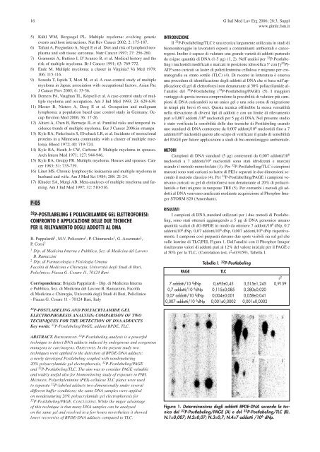
I campioni così preparati davano due spots visibili sia sul gel che<br />
sulle lastrine <strong>di</strong> TLC/PEI, Figura 1. Dall’analisi con il Phosphor Imager<br />
risultavano valori <strong>di</strong> addotti pari al 12% <strong>del</strong> valore iniziale per il PAGE e<br />
al 50% per la TLC, (Correlation test, r 2 =0,9159), Tabella I.<br />
Tabella I. 32 P-Postlabeling<br />
PAGE TLC<br />
r 2<br />
7 addotti/10 6 dNp 0,695±0,45 3,515±1,245 0,9159<br />
0,7 addotti/10 6 dNp 0,115±0,085 0,380±0,020<br />
0,07 addotti/10 6 dNp 0,004±0,001 0,058±0,041<br />
0,007 addotti/10 6 dNp 0,001±0,0002 0,001±0,0002<br />
Figura 1. Determinazione degli addotti BPDE-DNA secondo la tecnica<br />
<strong>del</strong> 32 P-Postlabeling/PAGE (A) e <strong>del</strong> 32 P-Postlabeling/TLC (B).<br />
N.1=0,007; N.2=0,07; N.3=0,7; N.4=7 addotti /10 6 dNp.