- Page 1 and 2:
Preface It has been more than a dec
- Page 3 and 4:
viii Contributors Björn P. Meij (5
- Page 5 and 6:
2 Chapter | 1 Concepts of Normality
- Page 7 and 8:
4 Chapter | 1 Concepts of Normality
- Page 9 and 10:
6 Chapter | 1 Concepts of Normality
- Page 11 and 12:
8 Chapter | 1 Concepts of Normality
- Page 13 and 14:
10 Chapter | 1 Concepts of Normalit
- Page 15 and 16:
12 Chapter | 1 Concepts of Normalit
- Page 17 and 18:
14 Chapter | 1 Concepts of Normalit
- Page 19 and 20:
16 Chapter | 1 Concepts of Normalit
- Page 21 and 22:
18 Chapter | 1 Concepts of Normalit
- Page 23 and 24:
20 Chapter | 1 Concepts of Normalit
- Page 25 and 26:
22 Chapter | 1 Concepts of Normalit
- Page 27 and 28:
24 Chapter | 1 Concepts of Normalit
- Page 29 and 30:
Chapter 2 Comparative Medical Genet
- Page 31 and 32:
I. Introduction 29 Green Red Green
- Page 33 and 34:
I. Introduction 31 A1 genetic map d
- Page 35 and 36:
I. Introduction 33 of genomes of va
- Page 38 and 39:
36 Chapter | 2 Comparative Medical
- Page 40 and 41:
38 Chapter | 2 Comparative Medical
- Page 42 and 43:
40 Chapter | 2 Comparative Medical
- Page 44 and 45:
42 Chapter | 2 Comparative Medical
- Page 46 and 47: 44 Chapter | 2 Comparative Medical
- Page 48 and 49: 46 Chapter | 3 Carbohydrate Metabol
- Page 50 and 51: 48 Chapter | 3 Carbohydrate Metabol
- Page 52 and 53: 50 Chapter | 3 Carbohydrate Metabol
- Page 54 and 55: 52 Chapter | 3 Carbohydrate Metabol
- Page 56 and 57: 54 Chapter | 3 Carbohydrate Metabol
- Page 58 and 59: 56 Chapter | 3 Carbohydrate Metabol
- Page 60 and 61: 58 Chapter | 3 Carbohydrate Metabol
- Page 62 and 63: 60 Chapter | 3 Carbohydrate Metabol
- Page 64 and 65: 62 Chapter | 3 Carbohydrate Metabol
- Page 66 and 67: 64 Chapter | 3 Carbohydrate Metabol
- Page 68 and 69: 66 Chapter | 3 Carbohydrate Metabol
- Page 71 and 72: IX. Disorders of Carbohydrate Metab
- Page 73 and 74: IX. Disorders of Carbohydrate Metab
- Page 75 and 76: IX. Disorders of Carbohydrate Metab
- Page 77 and 78: X. Disorders of Ruminants Associate
- Page 79 and 80: X. Disorders of Ruminants Associate
- Page 81 and 82: References 79 Kaneko , J. J. , Luic
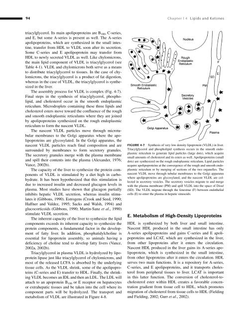
- Page 83 and 84: Chapter 4 Lipids and Ketones Michae
- Page 85 and 86: II. Long Chain Fatty Acids 83 FIGUR
- Page 87 and 88: II. Long Chain Fatty Acids 85 Plasm
- Page 89 and 90: IV. Phospholipids 87 The regulation
- Page 91 and 92: V. Cholesterol 89 V. CHOLESTEROL A.
- Page 93 and 94: VI. Lipoproteins 91 TABLE 4-1 Compo
- Page 95: VI. Lipoproteins 93 TABLE 4-2 Apoli
- Page 99 and 100: VIII. Ketogenesis and Ketosis 97 po
- Page 101 and 102: VIII. Ketogenesis and Ketosis 99 an
- Page 103 and 104: VIII. Ketogenesis and Ketosis 101 c
- Page 105 and 106: VIII. Ketogenesis and Ketosis 103 f
- Page 107 and 108: VIII. Ketogenesis and Ketosis 105 H
- Page 109 and 110: VIII. Ketogenesis and Ketosis 107 t
- Page 111 and 112: References 109 of free fatty acids
- Page 113 and 114: References 111 reducing equivalent
- Page 115 and 116: References 113 Oliver , M. F. , Kur
- Page 117 and 118: References 115 of peroxisomal β -o
- Page 119 and 120: 118 Chapter | 5 Proteins, Proteomic
- Page 121 and 122: 120 Chapter | 5 Proteins, Proteomic
- Page 123 and 124: 122 Chapter | 5 Proteins, Proteomic
- Page 125 and 126: 124 Chapter | 5 Proteins, Proteomic
- Page 127 and 128: 126 Chapter | 5 Proteins, Proteomic
- Page 129 and 130: 128 Chapter | 5 Proteins, Proteomic
- Page 131 and 132: 130 Chapter | 5 Proteins, Proteomic
- Page 133 and 134: 132 Chapter | 5 Proteins, Proteomic
- Page 135 and 136: 134 Chapter | 5 Proteins, Proteomic
- Page 137 and 138: 136 Chapter | 5 Proteins, Proteomic
- Page 139 and 140: 138 Chapter | 5 Proteins, Proteomic
- Page 141 and 142: 140 Chapter | 5 Proteins, Proteomic
- Page 143 and 144: 142 Chapter | 5 Proteins, Proteomic
- Page 145 and 146: 144 Chapter | 5 Proteins, Proteomic
- Page 147 and 148:
146 Chapter | 5 Proteins, Proteomic
- Page 149 and 150:
148 Chapter | 5 Proteins, Proteomic
- Page 151 and 152:
150 Chapter | 5 Proteins, Proteomic
- Page 153 and 154:
152 Chapter | 5 Proteins, Proteomic
- Page 155 and 156:
154 Chapter | 5 Proteins, Proteomic
- Page 157 and 158:
Chapter 6 Clinical Veterinary Immun
- Page 162 and 163:
162 Chapter | 6 Clinical Veterinary
- Page 164 and 165:
164 Chapter | 6 Clinical Veterinary
- Page 166 and 167:
166 Chapter | 6 Clinical Veterinary
- Page 168 and 169:
168 Chapter | 6 Clinical Veterinary
- Page 171:
VII. Modulation of the Immune Respo
- Page 174 and 175:
174 Chapter | 7 The Erythrocyte: Ph
- Page 176 and 177:
176 Chapter | 7 The Erythrocyte: Ph
- Page 178 and 179:
178 Chapter | 7 The Erythrocyte: Ph
- Page 180 and 181:
180 Chapter | 7 The Erythrocyte: Ph
- Page 182 and 183:
182 Chapter | 7 The Erythrocyte: Ph
- Page 184 and 185:
184 Chapter | 7 The Erythrocyte: Ph
- Page 186 and 187:
186 Chapter | 7 The Erythrocyte: Ph
- Page 188 and 189:
188 Chapter | 7 The Erythrocyte: Ph
- Page 190 and 191:
190 Chapter | 7 The Erythrocyte: Ph
- Page 192 and 193:
192 Chapter | 7 The Erythrocyte: Ph
- Page 194 and 195:
194 Chapter | 7 The Erythrocyte: Ph
- Page 196 and 197:
196 Chapter | 7 The Erythrocyte: Ph
- Page 198 and 199:
198 Chapter | 7 The Erythrocyte: Ph
- Page 200 and 201:
200 Chapter | 7 The Erythrocyte: Ph
- Page 202 and 203:
202 Chapter | 7 The Erythrocyte: Ph
- Page 204 and 205:
204 Chapter | 7 The Erythrocyte: Ph
- Page 206 and 207:
206 Chapter | 7 The Erythrocyte: Ph
- Page 208 and 209:
208 Chapter | 7 The Erythrocyte: Ph
- Page 210 and 211:
210 Chapter | 7 The Erythrocyte: Ph
- Page 212 and 213:
212 Chapter | 7 The Erythrocyte: Ph
- Page 214 and 215:
214 Chapter | 7 The Erythrocyte: Ph
- Page 216 and 217:
216 Chapter | 7 The Erythrocyte: Ph
- Page 218 and 219:
218 Chapter | 7 The Erythrocyte: Ph
- Page 220 and 221:
220 Chapter | 7 The Erythrocyte: Ph
- Page 222 and 223:
222 Chapter | 7 The Erythrocyte: Ph
- Page 224 and 225:
224 Chapter | 7 The Erythrocyte: Ph
- Page 226 and 227:
226 Chapter | 7 The Erythrocyte: Ph
- Page 228 and 229:
228 Chapter | 7 The Erythrocyte: Ph
- Page 230 and 231:
230 Chapter | 7 The Erythrocyte: Ph
- Page 232 and 233:
232 Chapter | 7 The Erythrocyte: Ph
- Page 234 and 235:
234 Chapter | 7 The Erythrocyte: Ph
- Page 236 and 237:
236 Chapter | 7 The Erythrocyte: Ph
- Page 238 and 239:
238 Chapter | 7 The Erythrocyte: Ph
- Page 240 and 241:
240 Chapter | 7 The Erythrocyte: Ph
- Page 242 and 243:
242 Chapter | 8 Porphyrins and the
- Page 244 and 245:
244 Chapter | 8 Porphyrins and the
- Page 246 and 247:
246 Chapter | 8 Porphyrins and the
- Page 248 and 249:
248 Chapter | 8 Porphyrins and the
- Page 250 and 251:
250 Chapter | 8 Porphyrins and the
- Page 252 and 253:
252 Chapter | 8 Porphyrins and the
- Page 254 and 255:
254 Chapter | 8 Porphyrins and the
- Page 256 and 257:
256 Chapter | 8 Porphyrins and the
- Page 258 and 259:
258 Chapter | 8 Porphyrins and the
- Page 260 and 261:
260 Chapter | 9 Iron Metabolism and
- Page 262 and 263:
262 Chapter | 9 Iron Metabolism and
- Page 264 and 265:
264 Chapter | 9 Iron Metabolism and
- Page 266 and 267:
266 Chapter | 9 Iron Metabolism and
- Page 268 and 269:
268 Chapter | 9 Iron Metabolism and
- Page 270 and 271:
270 Chapter | 9 Iron Metabolism and
- Page 272 and 273:
272 Chapter | 9 Iron Metabolism and
- Page 274 and 275:
274 Chapter | 9 Iron Metabolism and
- Page 276 and 277:
276 Chapter | 9 Iron Metabolism and
- Page 278 and 279:
278 Chapter | 9 Iron Metabolism and
- Page 280 and 281:
280 Chapter | 9 Iron Metabolism and
- Page 282 and 283:
282 Chapter | 9 Iron Metabolism and
- Page 284 and 285:
284 Chapter | 9 Iron Metabolism and
- Page 286 and 287:
Chapter 10 Hemostasis Patricia Gent
- Page 288 and 289:
II. Mechanisms of Hemostasis 289 FI
- Page 290 and 291:
II. Mechanisms of Hemostasis 291 TA
- Page 292 and 293:
II. Mechanisms of Hemostasis 293 gl
- Page 294 and 295:
II. Mechanisms of Hemostasis 295 th
- Page 296 and 297:
II. Mechanisms of Hemostasis 297 FI
- Page 298 and 299:
II. Mechanisms of Hemostasis 299 as
- Page 300 and 301:
II. Mechanisms of Hemostasis 301 th
- Page 302 and 303:
II. Mechanisms of Hemostasis 303 A
- Page 304 and 305:
II. Mechanisms of Hemostasis 305 co
- Page 306 and 307:
III. Laboratory Assessment of Hemos
- Page 308 and 309:
III. Laboratory Assessment of Hemos
- Page 310 and 311:
IV. Disorders of Hemostasis 311 TAB
- Page 312 and 313:
IV. Disorders of Hemostasis 313 of
- Page 314 and 315:
IV. Disorders of Hemostasis 315 b.
- Page 316 and 317:
IV. Disorders of Hemostasis 317 les
- Page 318 and 319:
IV. Disorders of Hemostasis 319 thr
- Page 320 and 321:
References 321 TABLE 10-9 Effects o
- Page 322 and 323:
References 323 Chao , W. , and Olso
- Page 324 and 325:
References 325 N. C. Jain , Eds.) ,
- Page 326 and 327:
References 327 Medved , L. , and Ni
- Page 328 and 329:
References 329 Russell , K. E. , an
- Page 330 and 331:
Chapter 11 Neutrophil Function Doug
- Page 332 and 333:
II. Neutrophil Functions 333 Select
- Page 334 and 335:
II. Neutrophil Functions 335 matrix
- Page 336 and 337:
II. Neutrophil Functions 337 the v-
- Page 338 and 339:
III. Virulence Factors Preventing N
- Page 340 and 341:
IV. Acquired Neutrophil Function De
- Page 342 and 343:
V. Neutrophil-Mediated Tissue Injur
- Page 344 and 345:
References 345 is thought to be due
- Page 346 and 347:
References 347 Goodger , W. J. , Fa
- Page 348 and 349:
References 349 Skoutelis , A. T. ,
- Page 350 and 351:
Chapter 12 Diagnostic Enzymology of
- Page 352 and 353:
III. Factors Affecting Serum Enzyme
- Page 354 and 355:
IV. Specific Enzymes 355 plays a ro
- Page 356 and 357:
IV. Specific Enzymes 357 reversible
- Page 358 and 359:
IV. Specific Enzymes 359 GGT is a m
- Page 360 and 361:
IV. Specific Enzymes 361 and early
- Page 362 and 363:
IV. Specific Enzymes 363 Studies of
- Page 364 and 365:
IV. Specific Enzymes 365 Serum CALP
- Page 366 and 367:
IV. Specific Enzymes 367 glycan lin
- Page 368 and 369:
IV. Specific Enzymes 369 Serum CK-M
- Page 370 and 371:
References 371 Arthur , J. R. ( 198
- Page 372 and 373:
References 373 Goldstein , D. J. ,
- Page 374 and 375:
References 375 Milan , J. L. , and
- Page 376 and 377:
References 377 Steiner , J. M. , an
- Page 378 and 379:
Chapter 13 Hepatic Function Bud C.
- Page 380 and 381:
III. Clinical Manifestations of Hep
- Page 382 and 383:
III. Clinical Manifestations of Hep
- Page 384 and 385:
III. Clinical Manifestations of Hep
- Page 386 and 387:
III. Clinical Manifestations of Hep
- Page 388 and 389:
IV. Laboratory Assessment of Hepati
- Page 390 and 391:
IV. Laboratory Assessment of Hepati
- Page 392 and 393:
IV. Laboratory Assessment of Hepati
- Page 394 and 395:
IV. Laboratory Assessment of Hepati
- Page 396 and 397:
IV. Laboratory Assessment of Hepati
- Page 398 and 399:
IV. Laboratory Assessment of Hepati
- Page 400 and 401:
IV. Laboratory Assessment of Hepati
- Page 402 and 403:
V. Overview and Conclusions 403 FIG
- Page 404 and 405:
References 405 extrahepatic bile du
- Page 406 and 407:
References 407 Haslewood , G. A. (
- Page 408 and 409:
References 409 Nilkumhang , P. , an
- Page 410 and 411:
References 411 Sutherland , R. J. ,
- Page 412 and 413:
Chapter 14 Gastrointestinal Functio
- Page 414 and 415:
III. Gastric Secretions 415 4 . Lip
- Page 416 and 417:
III. Gastric Secretions 417 FIGURE
- Page 418 and 419:
V. Exocrine Pancreatic Secretions 4
- Page 420 and 421:
V. Exocrine Pancreatic Secretions 4
- Page 422 and 423:
VII. Digestion and Absorption 423 c
- Page 424 and 425:
VII. Digestion and Absorption 425 T
- Page 426 and 427:
VII. Digestion and Absorption 427 h
- Page 428 and 429:
VII. Digestion and Absorption 429 i
- Page 430 and 431:
VII. Digestion and Absorption 431 L
- Page 432 and 433:
VIII. Disturbances of Gastrointesti
- Page 434 and 435:
VIII. Disturbances of Gastrointesti
- Page 436 and 437:
VIII. Disturbances of Gastrointesti
- Page 438 and 439:
VIII. Disturbances of Gastrointesti
- Page 440 and 441:
VIII. Disturbances of Gastrointesti
- Page 442 and 443:
VIII. Disturbances of Gastrointesti
- Page 444 and 445:
VIII. Disturbances of Gastrointesti
- Page 446 and 447:
References 447 characterized by red
- Page 448 and 449:
References 449 Brown , J. C. , Cook
- Page 450 and 451:
References 451 Glenert , J. , Jarnu
- Page 452 and 453:
References 453 Lecce , J. G. ( 1965
- Page 454 and 455:
References 455 intestinal absorptiv
- Page 456 and 457:
References 457 Webb , K. E. , Jr. (
- Page 458 and 459:
460 Chapter | 15 Skeletal Muscle Fu
- Page 460 and 461:
462 Chapter | 15 Skeletal Muscle Fu
- Page 462 and 463:
464 Chapter | 15 Skeletal Muscle Fu
- Page 464 and 465:
466 Chapter | 15 Skeletal Muscle Fu
- Page 466 and 467:
468 Chapter | 15 Skeletal Muscle Fu
- Page 468 and 469:
470 Chapter | 15 Skeletal Muscle Fu
- Page 470 and 471:
472 Chapter | 15 Skeletal Muscle Fu
- Page 472 and 473:
474 Chapter | 15 Skeletal Muscle Fu
- Page 474 and 475:
476 Chapter | 15 Skeletal Muscle Fu
- Page 476 and 477:
478 Chapter | 15 Skeletal Muscle Fu
- Page 478 and 479:
480 Chapter | 15 Skeletal Muscle Fu
- Page 480 and 481:
482 Chapter | 15 Skeletal Muscle Fu
- Page 482 and 483:
484 Chapter | 15 Skeletal Muscle Fu
- Page 484 and 485:
486 Chapter | 16 Kidney Function an
- Page 486 and 487:
488 Chapter | 16 Kidney Function an
- Page 488 and 489:
490 Chapter | 16 Kidney Function an
- Page 490 and 491:
492 Chapter | 16 Kidney Function an
- Page 492 and 493:
494 Chapter | 16 Kidney Function an
- Page 494 and 495:
496 Chapter | 16 Kidney Function an
- Page 496 and 497:
498 Chapter | 16 Kidney Function an
- Page 498 and 499:
500 Chapter | 16 Kidney Function an
- Page 500 and 501:
502 Chapter | 16 Kidney Function an
- Page 502 and 503:
504 Chapter | 16 Kidney Function an
- Page 504 and 505:
506 Chapter | 16 Kidney Function an
- Page 506 and 507:
508 Chapter | 16 Kidney Function an
- Page 508 and 509:
510 Chapter | 16 Kidney Function an
- Page 510 and 511:
512 Chapter | 16 Kidney Function an
- Page 512 and 513:
514 Chapter | 16 Kidney Function an
- Page 514 and 515:
516 Chapter | 16 Kidney Function an
- Page 516 and 517:
518 Chapter | 16 Kidney Function an
- Page 518 and 519:
520 Chapter | 16 Kidney Function an
- Page 520 and 521:
522 Chapter | 16 Kidney Function an
- Page 522 and 523:
524 Chapter | 16 Kidney Function an
- Page 524 and 525:
526 Chapter | 16 Kidney Function an
- Page 526 and 527:
528 Chapter | 16 Kidney Function an
- Page 528:
530 Chapter | 17 Fluid, Electrolyte
- Page 531:
V. Physiology of Acid-Base Balance
- Page 535 and 536:
V. Physiology of Acid-Base Balance
- Page 537 and 538:
V. Physiology of Acid-Base Balance
- Page 539 and 540:
V. Physiology of Acid-Base Balance
- Page 541 and 542:
VI. Evaluation of Imbalances 543 so
- Page 543 and 544:
VII. Clinical Features of Fluid and
- Page 545 and 546:
for the evaluation of acute fluid a
- Page 547 and 548:
VIII. Clinicopathological Indicator
- Page 549:
VIII. Clinicopathological Indicator
- Page 552 and 553:
554 Chapter | 17 Fluid, Electrolyte
- Page 554 and 555:
556 Chapter | 17 Fluid, Electrolyte
- Page 556 and 557:
558 Chapter | 17 Fluid, Electrolyte
- Page 558 and 559:
Chapter 18 Pituitary Function Jan A
- Page 560 and 561:
I. Hypothalamus-Pituitary System 56
- Page 562 and 563:
I. Hypothalamus-Pituitary System 56
- Page 564 and 565:
II. Anterior Lobe and Intermediate
- Page 566 and 567:
II. Anterior Lobe and Intermediate
- Page 568 and 569:
II. Anterior Lobe and Intermediate
- Page 570 and 571:
II. Anterior Lobe and Intermediate
- Page 572 and 573:
II. Anterior Lobe and Intermediate
- Page 574 and 575:
II. Anterior Lobe and Intermediate
- Page 576 and 577:
II. Anterior Lobe and Intermediate
- Page 578 and 579:
II. Anterior Lobe and Intermediate
- Page 580 and 581:
II. Anterior Lobe and Intermediate
- Page 582 and 583:
II. Anterior Lobe and Intermediate
- Page 584 and 585:
III. Neurohypophysis 587 Arginine v
- Page 586 and 587:
III. Neurohypophysis 589 in domesti
- Page 588 and 589:
IV. Assessment of Pituitary Functio
- Page 590 and 591:
References 593 of beta-endorphin an
- Page 592 and 593:
References 595 De Palo , E. F. , Ga
- Page 594 and 595:
References 597 vasopressin level an
- Page 596 and 597:
References 599 Liu , J. P. , Clarke
- Page 598 and 599:
Chapter | 7 Fluid, Electrolyte, and
- Page 600 and 601:
Chapter | 7 Fluid, Electrolyte, and
- Page 602 and 603:
Chapter 19 Adrenocortical Function
- Page 604 and 605:
II. Physiology of Adrenocortical Ho
- Page 606 and 607:
II. Physiology of Adrenocortical Ho
- Page 608 and 609:
II. Physiology of Adrenocortical Ho
- Page 610 and 611:
III. Adrenocortical Diseases 613 an
- Page 612 and 613:
IV. Assessment of Adrenocortical Fu
- Page 614 and 615:
IV. Assessment of Adrenocortical Fu
- Page 616 and 617:
References 619 b . Performance In t
- Page 618 and 619:
References 621 Ilett , K. F. , and
- Page 620 and 621:
Chapter 20 Thyroid Function J. Jerr
- Page 622 and 623:
V. Functions of the Thyroid Gland 6
- Page 624 and 625:
VII. Mechanism of Thyroid Hormone A
- Page 626 and 627:
X. Thyroid Function Tests 629 thyro
- Page 628 and 629:
X. Thyroid Function Tests 631 thyro
- Page 630 and 631:
References 633 the variations in TS
- Page 632 and 633:
Chapter 21 Clinical Reproductive En
- Page 634 and 635:
I. Introduction 637 e . Placental G
- Page 636 and 637:
II. Assay Methods 639 to another fo
- Page 638 and 639:
II. Assay Methods 641 compounds), n
- Page 640 and 641:
II. Assay Methods 643 and Nugent, 1
- Page 642 and 643:
III. Physiology of Reproductive Hor
- Page 644 and 645:
IV. Clinical Aspects of Reproductiv
- Page 646 and 647:
IV. Clinical Aspects of Reproductiv
- Page 648 and 649:
IV. Clinical Aspects of Reproductiv
- Page 650 and 651:
IV. Clinical Aspects of Reproductiv
- Page 652 and 653:
V. General Comments 655 F . Cat 1 .
- Page 654 and 655:
References 657 for storage at room
- Page 656 and 657:
References 659 Holtan , D. W. , Squ
- Page 658 and 659:
References 661 Sauer , M. J. , Foul
- Page 660 and 661:
Chapter 22 Trace Minerals Robert B.
- Page 662 and 663:
I. Introduction 665 The stability o
- Page 664 and 665:
II. Cobalt 667 TABLE 22-3 Potential
- Page 666 and 667:
III. Copper 669 methylmalonyl CoA m
- Page 668 and 669:
III. Copper 671 Tocopherol Mitochon
- Page 670 and 671:
III. Copper 673 acid on Cu absorpti
- Page 672 and 673:
III. Copper 675 pigs, deer, camels,
- Page 674 and 675:
IV. Manganese 677 IV . MANGANESE A
- Page 676 and 677:
IV. Manganese 679 lipid composition
- Page 678 and 679:
V. Molybdenum 681 FIGURE 22-5 Molyb
- Page 680 and 681:
VI. Selenium 683 and other lipid pe
- Page 682 and 683:
VI. Selenium 685 of Se deficiency o
- Page 684 and 685:
VII. Zinc 687 D . Zinc Metabolism,
- Page 686 and 687:
Although the syndrome described for
- Page 688 and 689:
References 691 Ensunsa , J. L. , Sy
- Page 690 and 691:
References 693 Subcommittee on Poul
- Page 692 and 693:
696 Chapter | 23 Vitamins FOOD RELE
- Page 694 and 695:
698 Chapter | 23 Vitamins A B C D R
- Page 696 and 697:
700 Chapter | 23 Vitamins Stellate
- Page 698 and 699:
702 Chapter | 23 Vitamins H CH H 3
- Page 700 and 701:
704 Chapter | 23 Vitamins plant tis
- Page 702 and 703:
706 Chapter | 23 Vitamins 1,25-(OH)
- Page 704 and 705:
708 Chapter | 23 Vitamins Infection
- Page 706 and 707:
710 Chapter | 23 Vitamins phylloqui
- Page 708 and 709:
712 Chapter | 23 Vitamins results i
- Page 710 and 711:
714 Chapter | 23 Vitamins NH 2 NH 2
- Page 712 and 713:
716 Chapter | 23 Vitamins b . Funct
- Page 714 and 715:
718 Chapter | 23 Vitamins occur in
- Page 716:
720 Chapter | 23 Vitamins Carnitine
- Page 719 and 720:
IV. Water-Soluble Vitamins 723 FIGU
- Page 721 and 722:
IV. Water-Soluble Vitamins 725 (i.e
- Page 723 and 724:
V. Vitamin-Like Compounds 727 4 . T
- Page 725 and 726:
References 729 Geraci , J. R. ( 197
- Page 727 and 728:
Chapter 24 Lysosomal Storage Diseas
- Page 729 and 730:
II. Lysosomal Storage Diseases (LSD
- Page 731 and 732:
II. Lysosomal Storage Diseases (LSD
- Page 733 and 734:
III. Pathogenesis 737 cellular orga
- Page 735 and 736:
V. Diagnosis 739 Weight in grams 80
- Page 737 and 738:
VI. Therapy 741 systems. Liver shou
- Page 739 and 740:
References 743 Broom , M. F. , Zhou
- Page 741 and 742:
References 745 Hubler , M. , Haskin
- Page 743 and 744:
References 747 Patterson , J. S. ,
- Page 745 and 746:
References 749 and Drinkwater , R.
- Page 747 and 748:
752 Chapter | 25 Tumor Markers comm
- Page 749 and 750:
754 Chapter | 25 Tumor Markers when
- Page 751 and 752:
756 Chapter | 25 Tumor Markers B. C
- Page 753 and 754:
758 Chapter | 25 Tumor Markers be d
- Page 755 and 756:
760 Chapter | 25 Tumor Markers the
- Page 757 and 758:
762 Chapter | 25 Tumor Markers Bost
- Page 759 and 760:
764 Chapter | 25 Tumor Markers Juba
- Page 761 and 762:
766 Chapter | 25 Tumor Markers 1,25
- Page 763 and 764:
Chapter 26 Cerebrospinal Fluid Will
- Page 765 and 766:
III. CSF Formation, Circulation, an
- Page 767 and 768:
IV. Cellular Composition of Normal
- Page 769 and 770:
V. Biochemical Constituents of Norm
- Page 771 and 772:
IV. Biochemical Constituents of Nor
- Page 773 and 774:
TABLE 26-7 Biochemical Constituents
- Page 775 and 776:
V. Biochemical Constituents of Norm
- Page 777 and 778:
V. Biochemical Constituents of Norm
- Page 779 and 780:
VI. CSF Collection and Analytical T
- Page 781 and 782:
VI. CSF Collection and Analytical T
- Page 783 and 784:
VI. CSF Collection and Analytical T
- Page 785 and 786:
VII. General Characteristics of CSF
- Page 787 and 788:
VII. General Characteristics of CSF
- Page 789 and 790:
VII. General Characteristics of CSF
- Page 791 and 792:
VII. General Characteristics of CSF
- Page 793 and 794:
VIII. Characteristics of CSF Associ
- Page 795 and 796:
VIII. Characteristics of CSF Associ
- Page 797 and 798:
VIII. Characteristics of CSF Associ
- Page 799 and 800:
VIII. Characteristics of CSF Associ
- Page 801 and 802:
VIII. Characteristics of CSF Associ
- Page 803 and 804:
References 809 M. , Petersen , A. ,
- Page 805 and 806:
References 811 deer (Odocoileus vir
- Page 807 and 808:
References 813 Hayward , R. A. , Sh
- Page 809 and 810:
References 815 Milhorat , T. H. ( 1
- Page 811 and 812:
References 817 amino acid concentra
- Page 813 and 814:
References 819 Waxman , F. J. , Cle
- Page 815 and 816:
822 Chapter | 27 Clinical Biochemis
- Page 817 and 818:
824 Chapter | 27 Clinical Biochemis
- Page 819 and 820:
826 Chapter | 27 Clinical Biochemis
- Page 821 and 822:
828 Chapter | 27 Clinical Biochemis
- Page 823 and 824:
830 Chapter | 27 Clinical Biochemis
- Page 825 and 826:
832 Chapter | 27 Clinical Biochemis
- Page 827 and 828:
834 Chapter | 27 Clinical Biochemis
- Page 829 and 830:
836 Chapter | 27 Clinical Biochemis
- Page 831 and 832:
Chapter 28 Avian Clinical Biochemis
- Page 833 and 834:
III. Starvation, Flight, and Postpr
- Page 835 and 836:
III. Starvation, Flight, and Postpr
- Page 837 and 838:
IV. Plasma Proteins 845 values can
- Page 839 and 840:
V. Renal Function 847 ASPERGILLOSIS
- Page 841 and 842:
V. Renal Function 849 from the fact
- Page 843 and 844:
Racing pigeon ALAT (IU/g tissue) Ra
- Page 845 and 846:
VI. Hepatobiliary Disease 853 Afric
- Page 847 and 848:
VI. Hepatobiliary Disease 855 TABLE
- Page 849 and 850:
VI. Hepatobiliary Disease 857 TABLE
- Page 851 and 852:
VIII. Calcium and Phosphorus: Metab
- Page 853 and 854:
VIII. Calcium and Phosphorus: Metab
- Page 855 and 856:
X. Exocrine Pancreatic Disease 863
- Page 857 and 858:
XI. Toxicology 865 Succinyl Co-A+Gl
- Page 859 and 860:
XII. Blood Coagulation 867 Coagulat
- Page 861 and 862:
References 869 Barthalmus , G. T. ,
- Page 863 and 864:
References 871 Lumeij , J. T. , and
- Page 865 and 866:
Appendix I SI Units The Systeme Int
- Page 867 and 868:
Appendix | I SI Units 875 TABLE E (
- Page 869 and 870:
Appendix III Temperature Correction
- Page 871 and 872:
Appendix V Temperature Conversions
- Page 873 and 874:
Appendix VII Conversions of Body We
- Page 875 and 876:
Appendix | VIII Blood Analyte Refer
- Page 877 and 878:
Appendix | VIII Blood Analyte Refer
- Page 879 and 880:
Appendix | VIII Blood Analyte Refer
- Page 881 and 882:
Appendix IX Blood Analyte Reference
- Page 883 and 884:
Appendix | IX Blood Analyte Referen
- Page 885 and 886:
Appendix | IX Blood Analyte Referen
- Page 887 and 888:
Appendix | IX Blood Analyte Referen
- Page 889 and 890:
Appendix | X Blood Analyte Referenc
- Page 891 and 892:
Appendix | XI Blood Analyte Referen
- Page 893 and 894:
Appendix XIII Cerebrospinal Fluid (
- Page 895 and 896:
Appendix XIV Cerebrospinal Fluid Re