- Page 1:
Salmonella enterica - biofilm forma
- Page 4 and 5:
Table of Contents Page Number 1.16.
- Page 6 and 7:
Table of Contents Page Number 4.1.2
- Page 8 and 9:
Table of Contents Page Number 6.8.4
- Page 10 and 11:
List of Abbreviations Table Page Nu
- Page 12 and 13:
List of Abbreviations List of Abbre
- Page 14 and 15:
Summary of Content Summary of Conte
- Page 16 and 17:
“To show your true ability is alw
- Page 18 and 19:
Acknowledgements ever met, Fiona th
- Page 20 and 21:
Acknowledgements This Ph.D. thesis
- Page 22 and 23:
Chapter 1 1.1. Salmonella Salmonell
- Page 24 and 25:
Chapter 1 may impose enormous costs
- Page 26 and 27:
Chapter 1 cracks or penetration of
- Page 28 and 29:
Chapter 1 detected by agglutination
- Page 30 and 31:
Chapter 1 become colonized with >10
- Page 32 and 33:
Chapter 1 antigens [z27],[z45] [1].
- Page 34 and 35:
Chapter 1 S. Agona has also been im
- Page 36 and 37:
Chapter 1 isolated from undercooked
- Page 38 and 39:
Chapter 1 The S. Typhimurium strain
- Page 40 and 41:
Chapter 1 ecosystem with the use of
- Page 42 and 43:
Chapter 1 [82]. Previous research h
- Page 44 and 45:
Chapter 1 to genes involved in flag
- Page 46 and 47:
Chapter 1 observation has also been
- Page 48 and 49:
Chapter 1 [119] and flow cell react
- Page 50 and 51:
Chapter 1 based disinfectant) the s
- Page 52 and 53:
Chapter 1 formation of two strains
- Page 54 and 55:
Chapter 1 shield the S. Agona cells
- Page 56 and 57:
Chapter 1 Key Objectives of this st
- Page 58 and 59:
Chapter 2 2. Abstract Food-borne pa
- Page 60 and 61:
Chapter 2 indicated that there may
- Page 62 and 63:
Chapter 2 taken from industrial set
- Page 64 and 65:
Chapter 2 2.1.4. CDC Biofilm Reacto
- Page 66 and 67:
Chapter 2 can be performed without
- Page 68 and 69:
Chapter 2 To summarise, previous au
- Page 70 and 71:
Chapter 2 2.3. Methods for examinin
- Page 72 and 73:
Chapter 2 and the biofilm formed th
- Page 74 and 75:
Chapter 2 2.4. Statistical Analysis
- Page 76 and 77:
Chapter 2 Table 2.1: Strain charact
- Page 78 and 79:
Chapter 2 XII. The gas port and med
- Page 80 and 81:
Chapter 2 II. Primary fixative cons
- Page 82 and 83:
Chapter 2 IX. Once the conditions w
- Page 84 and 85:
Chapter 2 Figure 2.1: Image of CBR
- Page 86 and 87:
Chapter 2 2.6. Results 2.6.1. Asses
- Page 88 and 89:
Chapter 2 2.6.3. SEM Analysis of su
- Page 90 and 91:
Chapter 2 Figure 2.6: Complete remo
- Page 92 and 93:
Chapter 2 2.6.5. Mean log 10 densit
- Page 94 and 95:
Chapter 2 2.6.6. Mean log 10 densit
- Page 96 and 97:
Chapter 2 Table 2.5: Mean log 10 de
- Page 98 and 99:
Chapter 2 The results displayed in
- Page 100 and 101:
Chapter 2 Table 2.7: Difference bet
- Page 102 and 103:
Chapter 2 Table 2.8: The mean log 1
- Page 104 and 105:
Chapter 2 Figure 2.7: Graph of the
- Page 106 and 107:
Chapter 2 also used “high” soni
- Page 108 and 109:
Chapter 2 2.8. Summary All 13 strai
- Page 110 and 111:
Chapter 3 3. Abstract It has been e
- Page 112 and 113:
Chapter 3 classified as persistent
- Page 114 and 115:
Chapter 3 was plated onto TSA for e
- Page 116 and 117:
Chapter 3 Figure 3.1: The mean log
- Page 118 and 119:
Chapter 3 Figure 3.3: SEM image of
- Page 120 and 121:
Chapter 3 Table 3.2: Difference bet
- Page 122 and 123:
Chapter 3 3.4.3. Intra-serovar vari
- Page 124 and 125:
Chapter 3 3.4.4. Strain variation i
- Page 126 and 127:
Chapter 3 3.4.5. The density of bio
- Page 128 and 129:
Chapter 3 3.5. Discussion As illust
- Page 130 and 131:
Chapter 3 As discussed in section 3
- Page 132 and 133:
Chapter 3 increased biofilm appeare
- Page 134 and 135:
Chapter 4 Examining the efficacy of
- Page 136 and 137:
Chapter 4 through laboratory based
- Page 138 and 139:
Chapter 4 acid and quaternary ammon
- Page 140 and 141:
Chapter 4 the surface the temperatu
- Page 142 and 143:
Chapter 4 availability of multiple
- Page 144 and 145:
Chapter 4 peracetic acid (100, 200,
- Page 146 and 147:
Chapter 4 As discussed previously,
- Page 148 and 149:
Chapter 4 Dey/Engley (Difco) neutra
- Page 150 and 151:
Chapter 4 sterilisation was achieve
- Page 152 and 153:
Chapter 4 IX. Aliquots of 100µl of
- Page 154 and 155:
Chapter 4 XV. XVI. XVII. XVIII. XIX
- Page 156 and 157: Chapter 4 4.3. Results 4.3.1. Suspe
- Page 158 and 159: Chapter 4 Table 4.3: Mean log 10 de
- Page 160 and 161: Chapter 4 Benzalkonium chloride was
- Page 162 and 163: Chapter 4 4.4. Discussion Suspensio
- Page 164 and 165: Chapter 4 effective at eliminating
- Page 166 and 167: Chapter 4 Surprisingly, Nguyen et a
- Page 168 and 169: Chapter 4 though a contact time of
- Page 170 and 171: Chapter 4 provide a more standardis
- Page 172 and 173: Chapter 4 hours instead of the stan
- Page 174 and 175: Chapter 5 5. Abstract Examining bio
- Page 176 and 177: Chapter 5 dry and rough (bdar) morp
- Page 178 and 179: Chapter 5 absence of curli and cell
- Page 180 and 181: Chapter 5 studies such as the micro
- Page 182 and 183: Chapter 5 discussed in chapter 4. T
- Page 184 and 185: Chapter 5 5.2. Methods 5.2.1. Colon
- Page 186 and 187: Chapter 5 XIII. XIV. XV. XVI. XVII.
- Page 188 and 189: Chapter 5 XVI. XVII. XVIII. XIX. XX
- Page 190 and 191: Chapter 5 5.3.2. Repeatability of m
- Page 192 and 193: Chapter 5 Figure 5.2: Stained biofi
- Page 194 and 195: Chapter 5 was formed at room temper
- Page 196 and 197: Chapter 5 Table 5.3: The density of
- Page 198 and 199: Chapter 5 5.3.4. The density of bio
- Page 200 and 201: Chapter 5 Table 5.6: The difference
- Page 202 and 203: Chapter 5 Table 5.7: The difference
- Page 204 and 205: Chapter 5 Table 5.8: Summary of ass
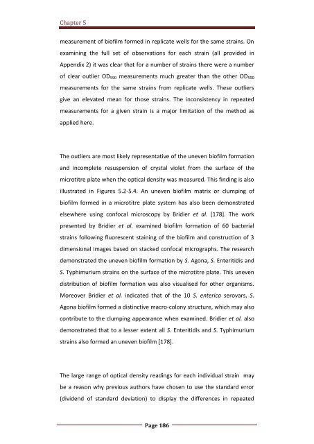
- Page 208 and 209: Chapter 5 measures of S. enterica b
- Page 210 and 211: Chapter 5 cells peaks [165, 191]. D
- Page 212 and 213: Chapter 5 5.5. Limitations of the s
- Page 214 and 215: Chapter 6 Discussion of S. enterica
- Page 216 and 217: Chapter 6 However, a number of auth
- Page 218 and 219: Chapter 6 It is of interest to exam
- Page 220 and 221: Chapter 6 were similar at 48 hours.
- Page 222 and 223: Chapter 6 attributable to a small n
- Page 224 and 225: Chapter 6 chosen as an appropriate
- Page 226 and 227: Chapter 6 undetermined. However, th
- Page 228 and 229: Chapter 6 reason for this may be du
- Page 230 and 231: Chapter 6 Moreover, based on the re
- Page 232 and 233: Chapter 6 responsible for increased
- Page 234 and 235: Chapter 6 not fully capture the ran
- Page 236 and 237: Chapter 6 However, the extent of bi
- Page 238 and 239: Chapter 6 6.10. Conclusions and rec
- Page 240 and 241: Chapter 6 studies may solve some of
- Page 242 and 243: Bibliography Bibliography Page 221
- Page 244 and 245: Bibliography 12. T. Takata, J.L., H
- Page 246 and 247: Bibliography 29. Barker, J., M. Nae
- Page 248 and 249: Bibliography 47. Threlfall, E.J., H
- Page 250 and 251: Bibliography 65. CDC. Investigation
- Page 252 and 253: Bibliography 83. Ledeboer, N., Frye
- Page 254 and 255: Bibliography 100. Chia, T., Goulter
- Page 256 and 257:
Bibliography 117. Buckingham-Meyer,
- Page 258 and 259:
Bibliography 133. European Committe
- Page 260 and 261:
Bibliography Supernatant of a Hafni
- Page 262 and 263:
Bibliography 165. Díez-García, M.
- Page 264 and 265:
Bibliography cultivation of Staphyl
- Page 266 and 267:
Bibliography Compounds. 2012. Appli
- Page 268 and 269:
Bibliography 215. Buck, et al., A q
- Page 270 and 271:
Bibliography Canadian Journal of Ps
- Page 272 and 273:
Appendix 1 Appendix 1— List of Re
- Page 274 and 275:
Appendix 1 Appendix 1 - List of Equ
- Page 276 and 277:
Appendix 2 Colour index High outlie
- Page 278 and 279:
Appendix 2 Colour index High outlie
- Page 280 and 281:
Appendix 2 Colour index High outlie
- Page 282 and 283:
Appendix 2 Colour index High outlie
- Page 284 and 285:
Appendix 2 Colour index High outlie
- Page 286 and 287:
Appendix 3 Appendix 3-Figure 2: PFG
- Page 288 and 289:
Appendix 4 Poster Presentation Envi


