LVR-Klinikum Düsseldorf Hospital of the Heinrich-Heine University ...
LVR-Klinikum Düsseldorf Hospital of the Heinrich-Heine University ...
LVR-Klinikum Düsseldorf Hospital of the Heinrich-Heine University ...

Create successful ePaper yourself
Turn your PDF publications into a flip-book with our unique Google optimized e-Paper software.
3.1.3.5 Gerontopsychiatry and dementia research<br />
Director: T. Supprian (from April 2005),<br />
C. Kretschmar (until April 2005)<br />
Coworkers: C. Lange-Asschenfeldt (from August 2005),<br />
R. Ihl (until April 2005)<br />
Early diagnosis and treatment <strong>of</strong> Alzheimer’s Dementia<br />
Director: T. Supprian, C. Lange-Asschenfeldt, as well as,<br />
for <strong>the</strong> Competence Network on Dementia: R. Ihl (until<br />
2005), C. Luckhaus (from 2005)<br />
Scientific assistants: T. Salamon, B. Höft, J. Szpak,<br />
B. Grass-Kapanke (until 2007), I. Blaeser (until 2008)<br />
Cooperation (external): H. Kessler, Department <strong>of</strong><br />
Psychiatry and Psycho<strong>the</strong>rapy, <strong>University</strong> <strong>Hospital</strong> Saarland,<br />
Homburg (Saar), K. Fast, Department <strong>of</strong> Psychiatry and<br />
Psycho<strong>the</strong>rapy, Ludwig-Maximilian <strong>University</strong>, Munich,<br />
as well as <strong>the</strong> Competence Network on Dementia<br />
(Spokesperson: W. Maier, Department <strong>of</strong> Psychiatry and<br />
Psycho<strong>the</strong>rapy <strong>of</strong> <strong>the</strong> <strong>University</strong> <strong>of</strong> Bonn).<br />
Financing: Funding from <strong>the</strong> Competence Network on<br />
Dementia, as well as departmental research budget<br />
In this research area, <strong>the</strong> projects started earlier in <strong>the</strong><br />
department were continued, and <strong>the</strong> memory outpatient unit<br />
(Director: C. Lange-Asschenfeldt) <strong>of</strong> <strong>the</strong> gerontopsychiatry<br />
institute outpatient clinic was used for research purposes.<br />
Worthy <strong>of</strong> note is <strong>the</strong> active participation in projects <strong>of</strong> <strong>the</strong><br />
Competence Network on Dementia. In addition to <strong>the</strong> larger<br />
projects, described in more detail below, in <strong>the</strong> context <strong>of</strong><br />
dissertations <strong>the</strong> working group also created an instrument<br />
for assessing dementia patients’ insight into <strong>the</strong>ir illness<br />
as well as <strong>the</strong> suitability <strong>of</strong> <strong>the</strong> Bielefeld Famous Faces<br />
Tests (BFFT) for <strong>the</strong> early diagnosis <strong>of</strong> depression and<br />
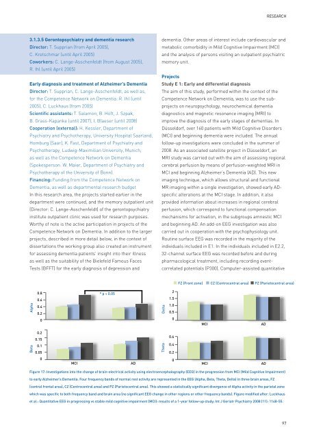
Alpha<br />
Beta<br />
0.8<br />
0.6<br />
0.4<br />
0.2<br />
0<br />
0.2<br />
0.15<br />
0.1<br />
0.05<br />
0<br />
* p < 0.05<br />
MCI AD<br />
MCI AD<br />
ReseaRch<br />
dementia. O<strong>the</strong>r areas <strong>of</strong> interest include cardiovascular and<br />
metabolic comorbidity in Mild Cognitive Impairment (MCI)<br />
and <strong>the</strong> analysis <strong>of</strong> persons visiting an outpatient psychiatric<br />
memory unit.<br />
Projects<br />
Study E 1: Early and differential diagnosis<br />
The aim <strong>of</strong> this study, performed within <strong>the</strong> context <strong>of</strong> <strong>the</strong><br />
Competence Network on Dementia, was to use <strong>the</strong> subprojects<br />
on neuropsychology, neurochemical dementia<br />
diagnostics and magnetic resonance imaging (MRI) to<br />
improve <strong>the</strong> diagnosis <strong>of</strong> <strong>the</strong> early stages <strong>of</strong> dementias. In<br />
<strong>Düsseldorf</strong>, over 160 patients with Mild Cognitive Disorders<br />
(MCI) and beginning dementia were included. The annual<br />
follow-up investigations were concluded in <strong>the</strong> summer <strong>of</strong><br />
2008. As an associated satellite project in <strong>Düsseldorf</strong>, an<br />
MRI study was carried out with <strong>the</strong> aim <strong>of</strong> assessing regional<br />
cerebral perfusion by means <strong>of</strong> perfusion-weighted MRI in<br />
MCI and beginning Alzheimer’s Dementia (AD). This new<br />
imaging technique, which allows structural and functional<br />
MR imaging within a single investigation, showed early ADspecific<br />
alterations at <strong>the</strong> MCI stage. In addition, it also<br />
provided information about increases in regional cerebral<br />
perfusion, which correspond to functional compensation<br />
mechanisms for activation, in <strong>the</strong> subgroups amnestic MCI<br />
and beginning AD. An add-on EEG investigation was also<br />
carried out in cooperation with <strong>the</strong> psychophysiology unit.<br />
Routine surface EEG was recorded in <strong>the</strong> majority <strong>of</strong> <strong>the</strong><br />
individuals included in E1. In <strong>the</strong> individuals included in E2.2,<br />
32-channel surface EEG was recorded before and during<br />
pharmacological treatment, including recording eventcorrelated<br />
potentials (P300). Computer-assisted quantitative<br />
Delta<br />
Theta<br />
2<br />
1.5<br />
1.0<br />
0.5<br />
0<br />
0.6<br />
0.4<br />
0.2<br />
0<br />
FZ (Front zone) CZ (Centrocentral area) PZ (Parietocentral area)<br />
MCI AD<br />
MCI AD<br />
Figure 17: Investigations into <strong>the</strong> change <strong>of</strong> brain-electrical activity using electroencephalography (EEG) in <strong>the</strong> progression from MCI (Mild Cognitive Impairment)<br />
to early Alzheimer’s Dementia. Four frequency bands <strong>of</strong> normal rest activity are represented in <strong>the</strong> EEG (Alpha, Beta, Theta, Delta) in three brain areas, FZ<br />
(central frontal area), CZ (Centrocentral area) and PZ (Parietocentral area). This showed a statistically significant divergence <strong>of</strong> Alpha activity in <strong>the</strong> parietal zone<br />
which was specific to both frequency band and brain area (no significant EEG change in o<strong>the</strong>r regions or o<strong>the</strong>r frequency bands). Figure modified after: Luckhaus<br />
et al.: Quantitative EEG in progressing vs stable mild cognitive impairment (MCI): results <strong>of</strong> a 1-year follow-up study; Int J Geriatr Psychiatry 2008 (11): 1148-55.<br />
97