Strato X User Manual - Image Works
Strato X User Manual - Image Works
Strato X User Manual - Image Works

Create successful ePaper yourself
Turn your PDF publications into a flip-book with our unique Google optimized e-Paper software.
USER'S MANUAL<br />
General instructions for use<br />
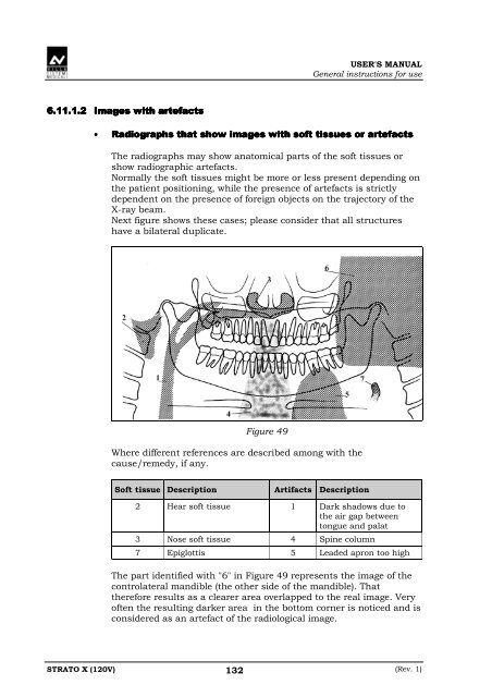
6.11.1.2 <strong>Image</strong>s with artefacts<br />
• Radiographs that show images with soft tissues or artefacts<br />
The radiographs may show anatomical parts of the soft tissues or<br />
show radiographic artefacts.<br />
Normally the soft tissues might be more or less present depending on<br />
the patient positioning, while the presence of artefacts is strictly<br />
dependent on the presence of foreign objects on the trajectory of the<br />
X-ray beam.<br />
Next figure shows these cases; please consider that all structures<br />
have a bilateral duplicate.<br />
Figure 49<br />
Where different references are described among with the<br />
cause/remedy, if any.<br />
Soft tissue Description Artifacts Description<br />
2 Hear soft tissue 1 Dark shadows due to<br />
the air gap between<br />
tongue and palat<br />
3 Nose soft tissue 4 Spine column<br />
7 Epiglottis 5 Leaded apron too high<br />
The part identified with "6" in Figure 49 represents the image of the<br />
controlateral mandible (the other side of the mandible). That<br />
therefore results as a clearer area overlapped to the real image. Very<br />
often the resulting darker area in the bottom corner is noticed and is<br />
considered as an artefact of the radiological image.<br />
STRATO X (120V) 132<br />
(Rev. 1)