Strato X User Manual - Image Works
Strato X User Manual - Image Works
Strato X User Manual - Image Works

Create successful ePaper yourself
Turn your PDF publications into a flip-book with our unique Google optimized e-Paper software.
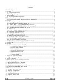
USER'S MANUAL<br />
General instructions for use<br />
6.7 IMPLANT examination<br />
The linear tomography examination (Implant) has been implemented to<br />
fulfil the emerging need of implant examination; in this way we make<br />
easiest the pre surgical planning mode and also to follow up the post<br />
surgical phase.<br />
Essential requirements for a tomographic examination in the surgical<br />
planning phase are to correctly determine the available space for implant;<br />
these requirements comprise the lingual and vestibular contour, bone<br />
thickness on the point of interest, position of the floor of the maxillary<br />
sinus or the distance between the alveolar crest and the upper board of<br />
the mandibular canal. All these structure are not adequately visualised<br />
using a standard panoramic or periapicals radiographs.<br />
Linear tomography technique make possible to answer to the above<br />
requirements with a constant magnification factor (+37%) both in vertical<br />
and horizontal direction, so making possible to perform measurements<br />
directly on the film. Additional advantage of linear tomography comes out<br />
from the dose reduction compared to the standard TC examination.<br />
<br />
NOTE:<br />
The presence of radio-opaque material close to the area under<br />
examination may generate artifacts which make difficult a good<br />
diagnosis.<br />
Implant examinations can be conducted with two different modalities,<br />
called reduced and complete implant respectively. First mode allows to<br />
obtain two images of the point of interest, while the second mode obtains<br />
four images; in both cases, examination is conducted in the same way.<br />
The images, having a dimensions of 5x7cm, are placed side by side on<br />
the same film. First image, the longitudinal allow a complete vision of the<br />
interested zone while the other are transversal images (1 or 3) of the<br />
selected point and allows to evaluate all the relevant dimensions (height<br />
and bone thickness).<br />
STRATO X (120V) 76<br />
(Rev. 1)