Zeitschrift für Rheumatologie – Supplement 1 - Deutsche ...
Zeitschrift für Rheumatologie – Supplement 1 - Deutsche ...
Zeitschrift für Rheumatologie – Supplement 1 - Deutsche ...
Erfolgreiche ePaper selbst erstellen
Machen Sie aus Ihren PDF Publikationen ein blätterbares Flipbook mit unserer einzigartigen Google optimierten e-Paper Software.
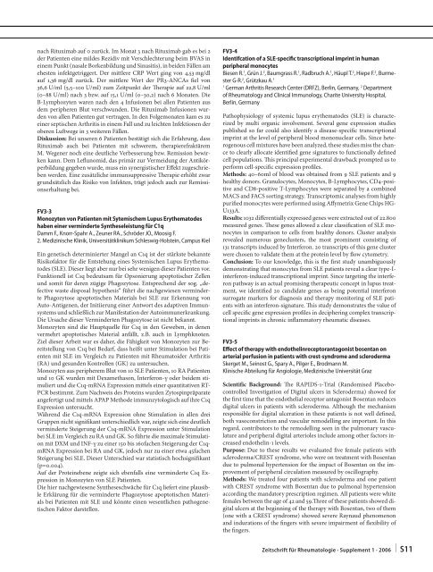
nach Rituximab auf 0 zurück. Im Monat 3 nach Rituximab gab es bei 2<br />
der Patienten eine mildes Rezidiv mit Verschlechterung beim BVAS in<br />
einem Punkt (nasale Borkenbildung und Sinusitis), in beiden Fällen am<br />
ehesten infektgetriggert. Der mittlere CRP Wert ging von 4,53 mg/dl<br />
auf 1,36 mg/dl zurück. Der mittlere Wert der PR3-ANCAs fi el von<br />
36,6 U/ml (5,5<strong>–</strong>100 U/ml) zum Zeitpunkt der Th erapie auf 22,8 U/ml<br />
(0<strong>–</strong>88 U/ml) nach 3 bzw. auf 15,1 U/ml (0<strong>–</strong>30,2) nach 6 Monaten. Die<br />
B-Lymphozyten waren nach den 4 Infusionen bei allen Patienten aus<br />
dem peripheren Blut verschwunden. Die Rituximab Infusionen wurden<br />
von allen Patienten gut vertragen. In den Folgemonaten kam es zu<br />
einer septischen Arthritis in einem Fall und zu leichten Infektionen der<br />
oberen Luft wege in 3 weiteren Fällen.<br />
Diskussion: Bei unseren 6 Patienten bestätigt sich die Erfahrung, dass<br />
Rituximab auch bei Patienten mit schwerem, therapierefraktärem<br />
M. Wegener noch eine deutliche Verbesserung bzw. Remission bewirken<br />
kann. Dem Lefl unomid, das primär zur Vermeidung der Antikörperbildung<br />
gegeben wurde, muss ein synergistischer Eff ekt zugeschrieben<br />
werden. Eine zusätzliche immunsuppressive Th erapie erhöht zwar<br />
grundsätzlich das Risiko von Infekten, trägt jedoch auch zur Remissionserhaltung<br />
bei.<br />
FV3-3<br />
Monozyten von Patienten mit Sytemischem Lupus Erythematodes<br />
haben einer verminderte Syntheseleistung <strong>für</strong> C1q<br />
Damm F., Knorr-Spahr A., Zeuner RA., Schröder JO., Moosig F.<br />
2. Medizinische Klinik, Universitätklinikum Schleswig-Holstein, Campus Kiel<br />
Ein genetisch determinierter Mangel an C1q ist der stärkste bekannte<br />
Risikofaktor <strong>für</strong> die Entstehung eines Systemischen Lupus Erythematodes<br />
(SLE). Dieser liegt aber nur bei sehr wenigen dieser Patienten vor.<br />
Funktionell ist C1q bedeutsam <strong>für</strong> Opsonierung apoptotischer Zellen<br />
und somit <strong>für</strong> deren zügige Phagozytose. Entsprechend der sog. „defective<br />
waste disposal hypothesis“ führt die nachgewiesen verminderte<br />
Phagozytose apoptotischen Materials bei SLE zur Erkennung von<br />
Auto-Antigenen, der Initiierung einer Antwort des adaptiven Immunsystems<br />
und schließlich zur Manifestation der Autoimmunerkrankung.<br />
Die Ursache dieser Verminderten Phagozytose ist nicht bekannt.<br />
Monozyten sind die Hauptquelle <strong>für</strong> C1q in den Geweben, in denen<br />
vermehrt apoptotisches Material anfällt, z.B. auch in Lymphknoten.<br />
Ziel dieser Arbeit war es daher, die Fähigkeit von Monozyten zur Bereitstellung<br />
von C1q bei Bedarf, dass heißt unter Stimulation bei Patienten<br />
mit SLE im Vergleich zu Patienten mit Rheumatoider Arthritis<br />
(RA) und gesunden Kontrollen (GK) zu untersuchen.<br />
Monozyten aus peripherem Blut von 10 SLE Patienten, 10 RA Patienten<br />
und 10 GK wurden mit Dexamethason, Interferon-γ oder beidem stimuliert<br />
und die C1q-mRNA Expression mittels einer quantitativen RT-<br />
PCR bestimmt. Zum Nachweis des Proteins wurden Zytospinpräparate<br />
angefertigt und mittels APAP Methode immunzytologisch auf ihre C1q<br />
Expression untersucht.<br />
Während die C1q-mRNA Expression ohne Stimulation in allen drei<br />
Gruppen nicht signifi kant unterschiedlich war, zeigte sich eine deutlich<br />
verminderte Steigerung der C1q-mRNA Expression unter Stimulation<br />
bei SLE im Vergleich zu RA und GK. So führte die maximale Stimulation<br />
mit DXM und INF-γ zu einer 150 bis 160fachen Steigerung der C1qmRNA<br />
Expression bei RA und GK, jedoch nur zu einer etwa 45fachen<br />
Steigerung bei SLE. Dieser Unterschied war statistisch hochsignifi kant<br />
(p=0.004).<br />
Auf der Proteinebene zeigte sich ebenfalls eine verminderte C1q Expression<br />
in Monozyten von SLE Patienten.<br />
Die hier nachgewiesene Syntheseschwäche <strong>für</strong> C1q liefert eine plausible<br />
Erklärung <strong>für</strong> die verminderte Phagozytose apoptotischen Materials<br />
bei Patienten mit SLE und könnte einen wesentlichen pathogenetischen<br />
Faktor darstellen.<br />
FV3-4<br />
Identifcation of a SLE-specifi c transcriptional imprint in human<br />
peripheral monocytes<br />
Biesen R. 1 , Grün J. 2 , Baumgrass R. 1 , Radbruch A. 1 , Häupl T. 2 , Hiepe F. 2 , Burmester<br />
G-R. 2 , Grützkau A. 1<br />
1 German Arthritis Research Center (DRFZ), Berlin, Germany, 2 Department<br />
of Rheumatology and Clinical Immunology, Charite University Hospital,<br />
Berlin, Germany<br />
Pathophysiology of systemic lupus erythematodes (SLE) is characterized<br />
by multi organic involvement. Several gene expression studies<br />
published so far could also identify a disease-specifi c transcriptional<br />
imprint at the level of peripheral blood mononuclear cells. Since heterogenous<br />
cell mixtures have been analyzed, these studies miss the chance<br />
to clearly allocate identifi ed gene signatures to functionally defi ned<br />
cell populations. Th is principal experimental drawback prompted us to<br />
perform cell-specifi c expression profi les.<br />
Methods: 40<strong>–</strong>60ml of blood was obtained from 9 SLE patients and 9<br />
healthy donors. Granulocytes, Monocytes, B-Lymphocytes, CD4-positive<br />
and CD8-positive T-Lymphocytes were separated by a combined<br />
MACS and FACS sorting strategy. Transcriptomic analyses from highly<br />
purifi ed monocytes were performed using Aff ymetrix Gene Chips HG-<br />
U133A.<br />
Results: 1032 diff erentially expressed genes were extracted out of 22.800<br />
measured genes. Th ese genes allowed a clear classifi cation of SLE monocytes<br />
in comparison to cells from healthy donors. Cluster analysis<br />
revealed numerous geneclusters, the most prominent consisting of<br />
131 trans cripts induced by Interferon. 20 transcripts of this gene cluster<br />
were chosen to validate them at the protein level by fl ow cytometry.<br />
Conclusion: To our knowledge, this is the fi rst study unambiguously<br />
demonstrating that monocytes from SLE patients reveal a clear type-Iinterferon-induced<br />
transcriptional imprint. Since targeting the interferon<br />
pathway is an actual promising therapeutic concept in lupus treatment,<br />
we identifi ed 20 candidate genes as being potential interferon<br />
surrogate markers for diagnosis and therapy monitoring of SLE patients<br />
with an interferon-signature. Th is study demonstrates the value of<br />
cell specifi c gene expression profi les in deciphering complex transcriptional<br />
imprints in chronic infl ammatory rheumatic diseases.<br />
FV3-5<br />
Eff ect of therapy with endothelinreceptorantagonist bosentan on<br />
arterial perfusion in patients with crest-syndrome and scleroderma<br />
Skerget M., Seinost G., Spary A., Pilger E., Brodmann M.<br />
Klinische Abteilung <strong>für</strong> Angiologie, Medizinische Universität Graz<br />
Scientific Background: Th e RAPIDS-1-Trial (Randomised Placebocontrolled<br />
Investigation of Digital ulcers in Scleroderma) showed for<br />
the fi rst time that the endothelial receptor antagonist Bosentan reduces<br />
digital ulcers in patients with scleroderma. Although the mechanism<br />
responsible for digital ulceration in these patients is not well defi ned,<br />
both vasoconstriction and vascular remodelling are important. In this<br />
regard, contributors to the remodelling seen in the pulmonary vasculature<br />
and peripheral digital arterioles include among other factors increased<br />
endothelin-1 levels.<br />
Purpose: Due to these results we evaluated fi ve female patients with<br />
scleroderma/CREST syndrome, who were on treatment with Bosentan<br />
due to pulmonal hypertension for the impact of Bosentan on the improvement<br />
of peripheral circulation measured by oscillography.<br />
Methods: We treated four patients with scleroderma and one patient<br />
with CREST syndrome with Bosentan due to pulmonal hypertension<br />
according the mandatory prescription regimen. All patients were white<br />
females between the age of 42 and 59.Th ree of these patients showed digital<br />
ulcers at the beginning of the therapy with Bosentan, two of them<br />
(one with a CREST syndrome) showed severe Raynaud phenomenon<br />
and indurations of the fi ngers with severe impairment of fl exibility of<br />
the fi ngers.<br />
<strong>Zeitschrift</strong> <strong>für</strong> <strong>Rheumatologie</strong> · <strong>Supplement</strong> 1 · 2006 | S11


