Smithsonian at the Poles: Contributions to International Polar
Smithsonian at the Poles: Contributions to International Polar
Smithsonian at the Poles: Contributions to International Polar

Create successful ePaper yourself
Turn your PDF publications into a flip-book with our unique Google optimized e-Paper software.
Many <strong>the</strong>ories have been hypo<strong>the</strong>sized <strong>to</strong> explain <strong>the</strong><br />
purpose and function of <strong>the</strong> erupted tusk. Proposed explan<strong>at</strong>ions<br />
include a weapon of aggression between males<br />
(Brown, 1868; Beddard, 1900; Lowe, 1906; Geist et al.,<br />
1960), a secondary sexual characteristic <strong>to</strong> establish social<br />
hierarchy among males (Scoresby, 1820; Hartwig,1874;<br />
Mansfi eld et al., 1975; Silverman and Dunbar, 1980),<br />
an instrument for breaking ice (Scoresby, 1820; Tomlin,<br />
1967), a spear for hunting (Vibe, 1950; Harrison and<br />
King, 1965; Bruemmer, 1993:64; Ellis, 1980), a bre<strong>at</strong>hing<br />
organ, a <strong>the</strong>rmal regul<strong>at</strong>or, a swimming rudder (Kingsley<br />
and Ramsay, 1988), a <strong>to</strong>ol for digging (Freuchen, 1935;<br />
Pederson, 1960; Newman, 1971), and an acoustic organ<br />
or sound probe (Best, 1981; Reeves and Mitchell, 1981).<br />
Examin<strong>at</strong>ion of tusk an<strong>at</strong>omy, his<strong>to</strong>logy, and biomechanics<br />
combined with traditional knowledge of Inuit elders<br />
and hunters has revealed fe<strong>at</strong>ures th<strong>at</strong> support a new<br />
sensory hypo<strong>the</strong>sis for tusk function (Nweeia et al., 2005).<br />
Narwhal tusk reaction and response <strong>to</strong> varying salinity gradients<br />
introduced during fi eld experiments support this.<br />
GROSS CRANIAL AND TOOTH ANATOMY<br />
Three narwhal head samples, obtained during legal<br />
Inuit harvests in 2003 and 2005 were examined by computerized<br />
axial <strong>to</strong>mography and magnetic resonance imaging<br />
and <strong>the</strong>n dissected. They included one adult male,<br />
one adult female, and one fetal specimen between four<br />
and six months in its development. The department of radiology<br />
<strong>at</strong> Johns Hopkins Hospital conducted computed<br />
<strong>to</strong>mography (CT) scans on all three specimens using a Siemens<br />
Medical Solutions SOMATOM Sens<strong>at</strong>ion Cardiac<br />
64. The scanner gener<strong>at</strong>ed 0.5-mm-thick slices on each of<br />
<strong>the</strong> three specimens. Original d<strong>at</strong>a from <strong>the</strong>se scans has<br />
been archived <strong>at</strong> <strong>the</strong> <strong>Smithsonian</strong> Institution. M<strong>at</strong>erialize<br />
Mimics 8.0 and Discreet 3D Studio Max 7 was used<br />
<strong>to</strong> cre<strong>at</strong>e digital 3-D models of narwhal dental an<strong>at</strong>omy.<br />
Magnetic resonance imaging (MRI) was also used <strong>to</strong> investig<strong>at</strong>e<br />
and visualize narwhal dental an<strong>at</strong>omy. Scientists<br />
<strong>at</strong> <strong>the</strong> N<strong>at</strong>ional Institutes of Health MRI Research Facility<br />
conducted MRI on <strong>the</strong> thawed narwhal heads. D<strong>at</strong>a<br />
from MRI assisted verifi c<strong>at</strong>ion of known cranial an<strong>at</strong>omy<br />
and enabled examin<strong>at</strong>ion of <strong>to</strong>oth vascul<strong>at</strong>ure. The narwhal<br />
heads were dissected <strong>at</strong> <strong>the</strong> Osteo-Prep Labor<strong>at</strong>ory<br />
<strong>at</strong> <strong>the</strong> <strong>Smithsonian</strong> Institution, and digital pho<strong>to</strong>graphs<br />
were taken <strong>to</strong> record an<strong>at</strong>omical landmarks and fe<strong>at</strong>ures<br />
of gross an<strong>at</strong>omy.<br />
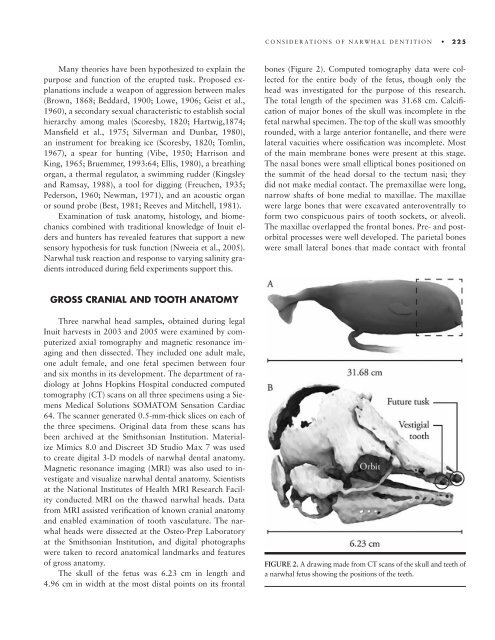
The skull of <strong>the</strong> fetus was 6.23 cm in length and<br />
4.96 cm in width <strong>at</strong> <strong>the</strong> most distal points on its frontal<br />
CONSIDERATIONS OF NARWHAL DENTITION 225<br />
bones (Figure 2). Computed <strong>to</strong>mography d<strong>at</strong>a were collected<br />
for <strong>the</strong> entire body of <strong>the</strong> fetus, though only <strong>the</strong><br />
head was investig<strong>at</strong>ed for <strong>the</strong> purpose of this research.<br />
The <strong>to</strong>tal length of <strong>the</strong> specimen was 31.68 cm. Calcifi -<br />
c<strong>at</strong>ion of major bones of <strong>the</strong> skull was incomplete in <strong>the</strong><br />
fetal narwhal specimen. The <strong>to</strong>p of <strong>the</strong> skull was smoothly<br />
rounded, with a large anterior fontanelle, and <strong>the</strong>re were<br />
l<strong>at</strong>eral vacuities where ossifi c<strong>at</strong>ion was incomplete. Most<br />
of <strong>the</strong> main membrane bones were present <strong>at</strong> this stage.<br />
The nasal bones were small elliptical bones positioned on<br />
<strong>the</strong> summit of <strong>the</strong> head dorsal <strong>to</strong> <strong>the</strong> tectum nasi; <strong>the</strong>y<br />
did not make medial contact. The premaxillae were long,<br />
narrow shafts of bone medial <strong>to</strong> maxillae. The maxillae<br />
were large bones th<strong>at</strong> were excav<strong>at</strong>ed anteroventrally <strong>to</strong><br />
form two conspicuous pairs of <strong>to</strong>oth sockets, or alveoli.<br />
The maxillae overlapped <strong>the</strong> frontal bones. Pre- and pos<strong>to</strong>rbital<br />
processes were well developed. The parietal bones<br />
were small l<strong>at</strong>eral bones th<strong>at</strong> made contact with frontal<br />
FIGURE 2. A drawing made from CT scans of <strong>the</strong> skull and teeth of<br />
a narwhal fetus showing <strong>the</strong> positions of <strong>the</strong> teeth.