Smithsonian at the Poles: Contributions to International Polar
Smithsonian at the Poles: Contributions to International Polar
Smithsonian at the Poles: Contributions to International Polar

Create successful ePaper yourself
Turn your PDF publications into a flip-book with our unique Google optimized e-Paper software.
238 SMITHSONIAN AT THE POLES / NWEEIA ET AL.<br />
as <strong>the</strong> algae deposits th<strong>at</strong> stain <strong>the</strong> surface would likely reappear<br />
if not continually removed. W<strong>at</strong>er turbulence alone<br />
would probably not account for removal of <strong>the</strong>se deposits<br />
from <strong>the</strong> tip and ridge areas of <strong>the</strong> tusk. The cleaned ridges<br />
are also smoo<strong>the</strong>d, indic<strong>at</strong>ing th<strong>at</strong> <strong>the</strong>y could be cleaned by<br />
physically rubbing against a surface such as ice. There have<br />
also been traditional knowledge descriptions of “tusking,”<br />
where males will g<strong>at</strong>her in small groups and rub tusks in<br />
a nonaggressive manner. Hunters clean harvested tusks by<br />
rubbing <strong>the</strong>m with sand <strong>to</strong> remove <strong>the</strong>se deposits. Perhaps<br />
tusks come in contact with sand and sediment when narwhal<br />
feed close <strong>to</strong> <strong>the</strong> bot<strong>to</strong>m.<br />
The cementum layer on <strong>the</strong> outer surface of <strong>the</strong> tusk<br />
is also a rare fe<strong>at</strong>ure for an erupted <strong>to</strong>oth. Cementum is<br />
generally found as a transitional layer between dentin and<br />
<strong>the</strong> periodontal ligaments th<strong>at</strong> hold teeth in<strong>to</strong> bone. These<br />
ligaments are able <strong>to</strong> <strong>at</strong>tach <strong>to</strong> <strong>the</strong> cementum with small<br />
fi bers, tying it <strong>to</strong> <strong>the</strong> surrounding bone. This appears <strong>to</strong> be<br />
consistent with <strong>the</strong> cementum observed in sections from<br />
<strong>the</strong> tusk base th<strong>at</strong> were <strong>at</strong>tached <strong>to</strong> segments of bone. In<br />
human teeth, however, if cementum becomes exposed <strong>to</strong><br />
<strong>the</strong> oral environment, it is rapidly worn away, exposing<br />
<strong>the</strong> root dentin. In <strong>the</strong> tusk, <strong>the</strong> cementum layer appears<br />
<strong>to</strong> remain intact, even after long exposure <strong>to</strong> <strong>the</strong> ocean<br />
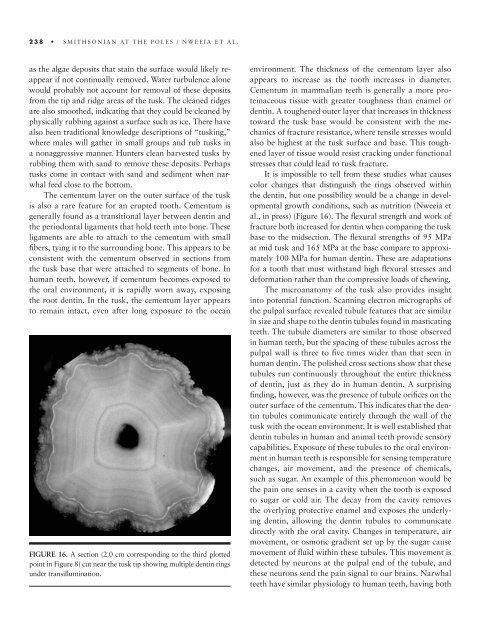
FIGURE 16. A section (2.0 cm corresponding <strong>to</strong> <strong>the</strong> third plotted<br />
point in Figure 8) cut near <strong>the</strong> tusk tip showing multiple dentin rings<br />
under transillumin<strong>at</strong>ion.<br />
environment. The thickness of <strong>the</strong> cementum layer also<br />
appears <strong>to</strong> increase as <strong>the</strong> <strong>to</strong>oth increases in diameter.<br />
Cementum in mammalian teeth is generally a more proteinaceous<br />
tissue with gre<strong>at</strong>er <strong>to</strong>ughness than enamel or<br />
dentin. A <strong>to</strong>ughened outer layer th<strong>at</strong> increases in thickness<br />
<strong>to</strong>ward <strong>the</strong> tusk base would be consistent with <strong>the</strong> mechanics<br />
of fracture resistance, where tensile stresses would<br />
also be highest <strong>at</strong> <strong>the</strong> tusk surface and base. This <strong>to</strong>ughened<br />
layer of tissue would resist cracking under functional<br />
stresses th<strong>at</strong> could lead <strong>to</strong> tusk fracture.<br />
It is impossible <strong>to</strong> tell from <strong>the</strong>se studies wh<strong>at</strong> causes<br />
color changes th<strong>at</strong> distinguish <strong>the</strong> rings observed within<br />
<strong>the</strong> dentin, but one possibility would be a change in developmental<br />
growth conditions, such as nutrition (Nweeia et<br />
al., in press) (Figure 16). The fl exural strength and work of<br />
fracture both increased for dentin when comparing <strong>the</strong> tusk<br />
base <strong>to</strong> <strong>the</strong> midsection. The fl exural strengths of 95 MPa<br />
<strong>at</strong> mid tusk and 165 MPa <strong>at</strong> <strong>the</strong> base compare <strong>to</strong> approxim<strong>at</strong>ely<br />
100 MPa for human dentin. These are adapt<strong>at</strong>ions<br />
for a <strong>to</strong>oth th<strong>at</strong> must withstand high fl exural stresses and<br />
deform<strong>at</strong>ion r<strong>at</strong>her than <strong>the</strong> compressive loads of chewing.<br />
The microan<strong>at</strong>omy of <strong>the</strong> tusk also provides insight<br />
in<strong>to</strong> potential function. Scanning electron micrographs of<br />
<strong>the</strong> pulpal surface revealed tubule fe<strong>at</strong>ures th<strong>at</strong> are similar<br />
in size and shape <strong>to</strong> <strong>the</strong> dentin tubules found in mastic<strong>at</strong>ing<br />
teeth. The tubule diameters are similar <strong>to</strong> those observed<br />
in human teeth, but <strong>the</strong> spacing of <strong>the</strong>se tubules across <strong>the</strong><br />
pulpal wall is three <strong>to</strong> fi ve times wider than th<strong>at</strong> seen in<br />
human dentin. The polished cross sections show th<strong>at</strong> <strong>the</strong>se<br />
tubules run continuously throughout <strong>the</strong> entire thickness<br />
of dentin, just as <strong>the</strong>y do in human dentin. A surprising<br />
fi nding, however, was <strong>the</strong> presence of tubule orifi ces on <strong>the</strong><br />
outer surface of <strong>the</strong> cementum. This indic<strong>at</strong>es th<strong>at</strong> <strong>the</strong> dentin<br />
tubules communic<strong>at</strong>e entirely through <strong>the</strong> wall of <strong>the</strong><br />
tusk with <strong>the</strong> ocean environment. It is well established th<strong>at</strong><br />
dentin tubules in human and animal teeth provide sensory<br />
capabilities. Exposure of <strong>the</strong>se tubules <strong>to</strong> <strong>the</strong> oral environment<br />
in human teeth is responsible for sensing temper<strong>at</strong>ure<br />
changes, air movement, and <strong>the</strong> presence of chemicals,<br />
such as sugar. An example of this phenomenon would be<br />
<strong>the</strong> pain one senses in a cavity when <strong>the</strong> <strong>to</strong>oth is exposed<br />
<strong>to</strong> sugar or cold air. The decay from <strong>the</strong> cavity removes<br />
<strong>the</strong> overlying protective enamel and exposes <strong>the</strong> underlying<br />
dentin, allowing <strong>the</strong> dentin tubules <strong>to</strong> communic<strong>at</strong>e<br />
directly with <strong>the</strong> oral cavity. Changes in temper<strong>at</strong>ure, air<br />
movement, or osmotic gradient set up by <strong>the</strong> sugar cause<br />
movement of fl uid within <strong>the</strong>se tubules. This movement is<br />
detected by neurons <strong>at</strong> <strong>the</strong> pulpal end of <strong>the</strong> tubule, and<br />
<strong>the</strong>se neurons send <strong>the</strong> pain signal <strong>to</strong> our brains. Narwhal<br />
teeth have similar physiology <strong>to</strong> human teeth, having both