

Reproduction in Domestic Animals
Reproduction in Domestic Animals
Reproduction in Domestic Animals
- No tags were found...

You also want an ePaper? Increase the reach of your titles
YUMPU automatically turns print PDFs into web optimized ePapers that Google loves.
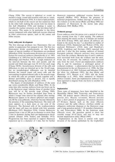
152 H L<strong>in</strong>debergChang 1950). The oocyte is spherical or ovoid; itsnucleus is large, round and eccentric with one or, rarely,two nucleoli (Rob<strong>in</strong>son 1918). It is rich <strong>in</strong> lipid particles,which <strong>in</strong> one-half of the oocyte are less numerous than<strong>in</strong> the other half, mak<strong>in</strong>g the oocyte look less dense <strong>in</strong>this half (Hamilton 1934) and mak<strong>in</strong>g it easier tovisualize the nuclear structures on the less dense half ofthe oocyte. This seems to be particular to the ferretoocyte compared with other lipid-rich oocytes observed<strong>in</strong> other carnivorous species, such as the can<strong>in</strong>e andfel<strong>in</strong>e oocytes.Early embryonic developmentThe first cleavage produces two blastomeres that aresimilar <strong>in</strong> appearance but unequal <strong>in</strong> size. The first twoblastomeres divide asynchronously, so that furtherstages of uneven numbers of blastomeres are produced(Hamilton 1934). Asynchronous divisions are a commonfeature for carnivore embryos (Amstislavsky et al. 1993;L<strong>in</strong>deberg et al. 1993) but not for embryos of rum<strong>in</strong>ants(Betteridge and Fle´ chon 1988). A rough estimation ofthe <strong>in</strong>tervals between the first and second, and thesecond and third cleavages was 10–16 h for each stage(Chang 1965b). Asynchronous division of the cells anddifferences <strong>in</strong> sizes are detected up to the 16-cell stage,but no morphological differences can be detectedbetween the central (<strong>in</strong>ner cell mass) cells and thesurround<strong>in</strong>g (trophoectoderm) cells at the morula stage,<strong>in</strong> which the cells are grouped closely together and adist<strong>in</strong>ct perivitell<strong>in</strong>e space is present. Fat is equallydistributed among the cells (Hamilton 1934).The degree of development of embryos of the sameage varies considerably (Rider and Heap 1986). At thesame time after mat<strong>in</strong>g, embryos from one ferret can beat the blastocyst stage, whereas those of another ferretmay still be at the morula stage (Chang 1969).Blastocysts may be further developed than those <strong>in</strong>which a longer time <strong>in</strong>terval has elapsed after mat<strong>in</strong>g.Blastocysts appear to be almost spherical and completelyfill the zona pellucida, which has become th<strong>in</strong>ned bythe presence of the expand<strong>in</strong>g blastocyst. The <strong>in</strong>ner cellmass appears as a dark mass at one pole of theblastocyst. The flattened cells of the trophoectodermand the <strong>in</strong>ner cell mass are dotted with fatty granules(Rob<strong>in</strong>son 1925; Hamilton 1934). Uter<strong>in</strong>e blastocystsexpand from a size of 200 lm <strong>in</strong> diameter to more than2 mm <strong>in</strong> diameter dur<strong>in</strong>g their pre-implantation development(Daniel 1970; Enders and Schlafke 1972).Progesterone has been reported to support blastocystexpansion up to a diameter of 1 mm, but for furtherblastocyst expansion additional ovarian factors arerequired (McRae 1992). Without the presence ofmaternal progesterone, neither cleavage of embryos <strong>in</strong>the oviducts or <strong>in</strong> the uterus (Rider and Heap 1986) norexpansion of blastocysts <strong>in</strong> the uterus take place(Buchanan 1969; McRae 1992).Oviductal passageFerret embryos enter the uterus over a period of severaldays start<strong>in</strong>g from day 5 after mat<strong>in</strong>g. The embryosstart enter<strong>in</strong>g the uterus on day 4, if females are treatedwith hCG (Chang 1969), and almost all embryos arefound <strong>in</strong> the uterus by day 7 as described <strong>in</strong> studies ofRob<strong>in</strong>son (1918), Hammond and Walton (1934), whoanalyzed Rob<strong>in</strong>son’s (1918) data and Hamilton’s(1934). L<strong>in</strong>deberg and Ja¨ rv<strong>in</strong>en (2003) reported thatuntil day 5 after the first mat<strong>in</strong>g, embryos of farmedEuropean polecats (Mustela putorius) were recoveredonly from the oviducts. Between days 6 and 9, embryoswere recovered both from the oviducts and the uteri.From day 10 onwards, the embryos were recoveredonly from the uteri. Ferret pre-implantation embryos(Fig. 1a and b) experience a prolonged period ofoviductal residence – a phenomenon that has also beendemonstrated <strong>in</strong> the cat (Swanson et al. 1994), the bluefox (Valtonen et al. 1985; Valtonen and Jalkanen 1993),the silver fox (Jalkanen 1992), the dog (Holst andPhemister 1971; Renton et al. 1991) and the horse(Betteridge et al. 1982). After unilateral or bilateralembryo transfer, ferret pre-implantation embryos havebeen reported to migrate from one horn to another (Liet al. 2006).ImplantationThree types of pregnancy have been identified <strong>in</strong> theMustelidae (Mead 1989; Ternovsky and Ternovskaya1994). All polecat species (Mustela putorius, Mustelaputorius furo, Mustela eversmanni, Mustela lutreola)have a short period of pregnancy of constant duration(37–44 days). Species such as the stoat (Mustelaerm<strong>in</strong>ea) and the sable (Martes zibell<strong>in</strong>a) exhibit anobligatory embryonic diapause at the blastocyst stageand a long gestation period (7–10 months) (Amstislavskyand Ternovskaya 2000). In the American m<strong>in</strong>k(Mustela vison), the gestation period is short butvariable (range 45–61 days), and may or may not<strong>in</strong>clude implantation delay (Mead 1989).Implantation <strong>in</strong> the ferret is central with rapid<strong>in</strong>vasion of the uter<strong>in</strong>e epithelium by the trophoblast(a)(b)Fig. 1. (a) Freshly flushed day-9blastocysts; (b) Frozen-thawedday-9 blastocysts <strong>in</strong> the farmedEuropean polecatÓ 2008 The Author. Journal compilation Ó 2008 Blackwell Verlag


